Bone Age Estimation Chart
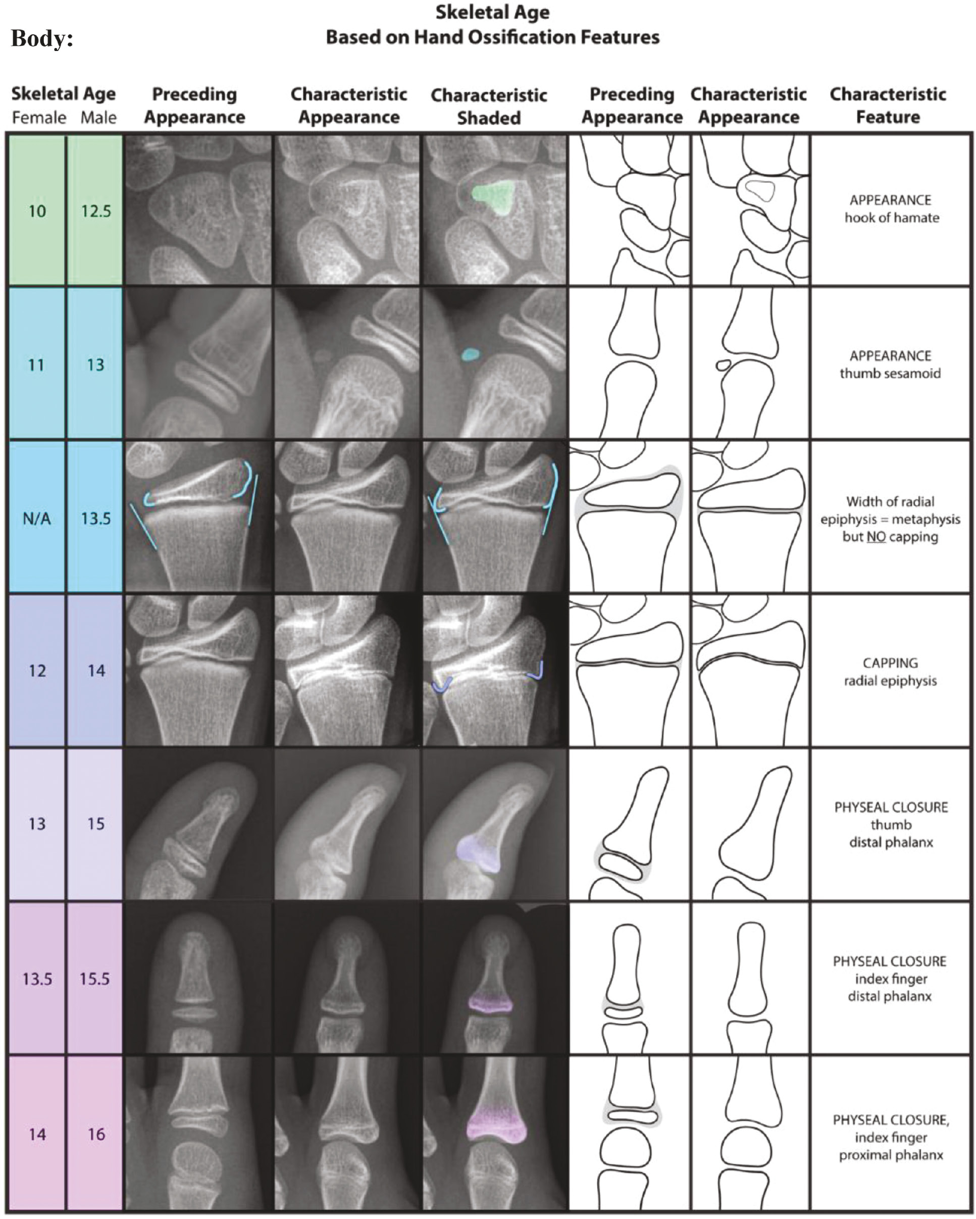
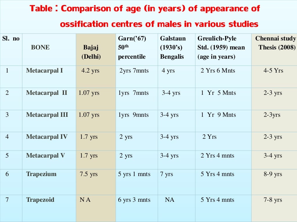
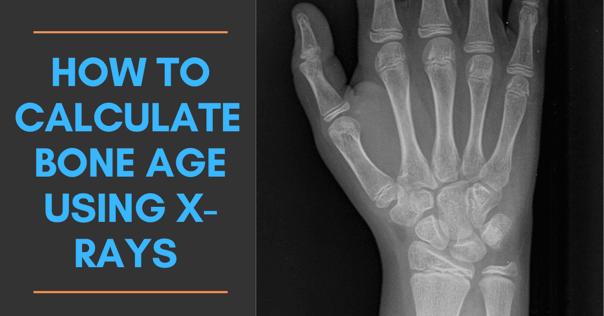
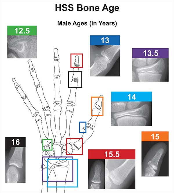
Bone Age Estimation Chart - Web the radiograph is used in conjunction with clinically validated nomograms to determine skeletal age. Bone age continues to be a valuable tool in assessing children’s health. Web pediatricians have relied on methods for determining skeletal maturation for >75 years. The greulich and pyle method makes use of a standard bone age atlas that the reporter can compare their image to and make an estimation of bone age. Review the clinical relevance of determining bone age. Patients with delayed bone age, patients with bone age appropriate to chronological age,. Tanner and whitehouse’ and greulich and pyle’s are some well known ones. Web by evaluating the data obtained from bone age in the clinical setting, it is possible to distinguish three main groups of subjects: Web a single reference guide that provides an anatomically comprehensive overview to the principles associated with skeletal age estimation, bringing an array of current validated bone age determination methods together, with easily referenced visual presentations and elements of consistency throughout the systems, may be of great utility to pediatr. Web both main methods of bone age assessment require a left hand and wrist radiograph. Web this chapter describes the anatomical and physiologic principles underlying bone age, factors that generally lead to delayed or advanced bone ages, methods of bone age determination, common clinical scenarios in which bone age assessment may be useful, and the use of bone age for height prediction. Explain the role of the interprofessional team in determining bone age and how. Bone age continues to be a valuable tool in assessing children’s health. Web both main methods of bone age assessment require a left hand and wrist radiograph. Explain the role of the interprofessional team in determining bone age and how it can lead to improved outcomes. Web the radiograph is used in conjunction with clinically validated nomograms to determine skeletal. Web this chapter describes the anatomical and physiologic principles underlying bone age, factors that generally lead to delayed or advanced bone ages, methods of bone age determination, common clinical scenarios in which bone age assessment may be useful, and the use of bone age for height prediction. The greulich and pyle method makes use of a standard bone age atlas. It forms an important part of the diagnostic and management pathway in children with growth and endocrine disorders. Web by the age of 18 years, bone age cannot be computed from hand & wrist radiographs, therefore the medial end of the clavicle is used for bone age calculation in individuals aged 18—22 years. Hernández method) or body weight (up to. Web by the age of 18 years, bone age cannot be computed from hand & wrist radiographs, therefore the medial end of the clavicle is used for bone age calculation in individuals aged 18—22 years. Explain the role of the interprofessional team in determining bone age and how it can lead to improved outcomes. Web a single reference guide that. Web by evaluating the data obtained from bone age in the clinical setting, it is possible to distinguish three main groups of subjects: Bone age continues to be a valuable tool in assessing children’s health. Web this chapter describes the anatomical and physiologic principles underlying bone age, factors that generally lead to delayed or advanced bone ages, methods of bone. Explain the role of the interprofessional team in determining bone age and how it can lead to improved outcomes. Ct visualization of the clavicle has been extensively studied but requires a high dose of radiation. Describe the indications for determining bone age. Web both main methods of bone age assessment require a left hand and wrist radiograph. Web this chapter. Web pediatricians have relied on methods for determining skeletal maturation for >75 years. The greulich and pyle method makes use of a standard bone age atlas that the reporter can compare their image to and make an estimation of bone age. Web the bone age radiograph of the hand and wrist is a commonly performed examination to determine the radiographic. Web by evaluating the data obtained from bone age in the clinical setting, it is possible to distinguish three main groups of subjects: Web a single reference guide that provides an anatomically comprehensive overview to the principles associated with skeletal age estimation, bringing an array of current validated bone age determination methods together, with easily referenced visual presentations and elements. Web bone age assessment (baa) is crucial in the evaluation of endocrine disorders and in the prediction of adult height when hormone therapy is the treatment [ 1 ]. Web the radiograph is used in conjunction with clinically validated nomograms to determine skeletal age. Several other bone age assessment methods have been developed, including ultrasonographic, computerized, and magnetic resonance (mr). Bone age assessment plays a crucial role in pediatric radiology, helping to determine a child’s level of maturation and growth potential. Describe the indications for determining bone age. Methods and analysis this is a prospective, observational study. Web pediatricians have relied on methods for determining skeletal maturation for >75 years. Web a single reference guide that provides an anatomically comprehensive overview to the principles associated with skeletal age estimation, bringing an array of current validated bone age determination methods together, with easily referenced visual presentations and elements of consistency throughout the systems, may be of great utility to pediatr. Explain the role of the interprofessional team in determining bone age and how it can lead to improved outcomes. Bone age continues to be a valuable tool in assessing children’s health. Web by the age of 18 years, bone age cannot be computed from hand & wrist radiographs, therefore the medial end of the clavicle is used for bone age calculation in individuals aged 18—22 years. Several other bone age assessment methods have been developed, including ultrasonographic, computerized, and magnetic resonance (mr) imaging methods. Tanner and whitehouse’ and greulich and pyle’s are some well known ones. Web the bone age radiograph of the hand and wrist is a commonly performed examination to determine the radiographic age of the patient via the assessment of growth centers. Short stature / poor growth, e.g. The greulich and pyle method makes use of a standard bone age atlas that the reporter can compare their image to and make an estimation of bone age. Patients with delayed bone age, patients with bone age appropriate to chronological age,. Web bone age assessment (baa) is crucial in the evaluation of endocrine disorders and in the prediction of adult height when hormone therapy is the treatment [ 1 ]. Web the radiograph is used in conjunction with clinically validated nomograms to determine skeletal age.
Bone Age Estimation Chart Wrist

Example of bone age determination. A girl with chronological age of 12

Age estimation by bones

UtahRad Bone age determination in infants

Growth chart of our patient (Δ height for bone age). Download

Height growth chart, showing progression of chronological age and bone

Skeletal Age Radiology Key

Calculating Bone Age Using Xrays [Includes AIbased Automated Tools

Skeletal maturity chart for boys comparing bone age and chronologic

A New Validated Shorthand Method for Determining Bone Age
Hernández Method) Or Body Weight (Up To 1 Year Of Age;
Ct Visualization Of The Clavicle Has Been Extensively Studied But Requires A High Dose Of Radiation.
It Forms An Important Part Of The Diagnostic And Management Pathway In Children With Growth And Endocrine Disorders.
Depending On The Method, Either Age (Up To 2 Years Of Age;
Related Post: