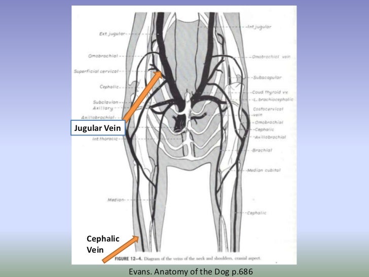
Cephalic Vein Dog Blood Draw
Cephalic Vein Dog Blood Draw - Otherwise, one quick and controlled motion is often enough to enter the vein. It begins on the mediopalmar surface of the carpus where it is a continuation of the radial vein. Blood appears in the needle hub when the needle enters the vein. Typically, a 22g needle (blue needle) works for most blood draws. Collection of blood for analysis. Web blood samples are commonly obtained from the jugular, cephalic or lateral saphenous veins; Once blood collected, let off vein, before the needle removed, still hold onto leg, then cover puncture site when needle out. Contributed by nichola gaither from animal hospital of statesville. Roth dr, and bürki k. To use a vein close to the surface of the body; Web the cephalic vein is wiped with alcohol. It is the only large superficial vein of the thoracic limb. That might include the cephalic vein. Web the cephalic vein is relatively large and often visible through the skin, and can be an attractive candidate for healthcare practitioners to draw blood through venipuncture. The simple answer is from the vein. To be able to gently withdraw a suitable volume of blood (to minimise damage to blood cells). Collection of blood for analysis. First, the needle needs to enter the patient’s skin bevel up. Web the lateral saphenous vein in dogs is an ideal spot for quick blood draws. Medium to larger dog cephalic or saphenous vein. Roth dr, and bürki k. Collection of blood for analysis. Otherwise, one quick and controlled motion is often enough to enter the vein. Second, we want to break the seal on the syringe. Web common blood draw locations for dogs are jugular veins, which run on either side of the windpipe. Peripheral veins, eg cephalic and saphenous, often detrimental → slow blood flow → sample artefacts (hemolysis and microclots.) uses. Web the lateral saphenous vein in dogs is an ideal spot for quick blood draws. The number of attempts to obtain blood should be minimized to a maximum of three needle sticks for each sample. Contributed by nichola gaither from animal. While facing your dog, have them shake, then briefly cup their foreleg with one hand and treat. Dogs can either be restrained manually or using a sling. Web the lateral saphenous vein in dogs is an ideal spot for quick blood draws. Web good sample collection technique is vital to obtain a blood sample of adequate quality for analysis. Web. Next, use your dominant hand to hold the syringe and swiftly insert the needle into the “vein.”. Web in this episode of breeders hacks‼️we talk about new tips and tricks to be easily able to draw your dogs blood from even at home‼️_____. Web the lateral saphenous vein in dogs is an ideal spot for quick blood draws. The rvn. It begins on the mediopalmar surface of the carpus where it is a continuation of the radial vein. It is more convenient to collect (draw) blood from a dog’s cephalic or saphenous veins than the jugular vein. Alexander street is an imprint of proquest that promotes teaching, research, and learning across music, counseling, history, anthropology, drama, film, and more. Web. No more than eight blood samples (four from either leg) should be taken in any 24 hour period. Web the lateral saphenous vein in dogs is an ideal spot for quick blood draws. The medial saphenous vein can also be used, and in general the use of the jugular vein is recommended. The simple answer is from the vein. Web. Web good sample collection technique is vital to obtain a blood sample of adequate quality for analysis. Dogs can either be restrained manually or using a sling. Web the lateral saphenous vein in dogs is an ideal spot for quick blood draws. Alexander street is an imprint of proquest that promotes teaching, research, and learning across music, counseling, history, anthropology,. No more than eight blood samples (four from either leg) should be taken in any 24 hour period. It is the only large superficial vein of the thoracic limb. If your dog is trained to shake, you’re halfway to the other part of cephalic blood draw training: The number of attempts to obtain blood should be minimized to a maximum. Approximately 0.5 to 0.75 cm of the needle is placed into the vein. We are going to show you how to draw from the cephalic vein. Dogs can either be restrained manually or using a sling. Web dovelewis technician kara prater, cvt, demonstrates the steps, equipment, and restraint needed for a cephalic vein blood draw on a canine patient.join this c. Blood appears in the needle hub when the needle enters the vein. Contributed by nichola gaither from animal hospital of statesville. No more than eight blood samples (four from either leg) should be taken in any 24 hour period. Blood collection from the sublingual vein in mice and hamsters:. To be able to gently withdraw a suitable volume of blood (to minimise damage to blood cells). Web the cephalic vein is wiped with alcohol. The medial saphenous vein can also be used, and in general the use of the jugular vein is recommended. The rvn or svn should be proficient in the technique for optimal sample procurement to prevent problems with the sample that may impact. Medium to larger dog cephalic or saphenous vein. Twisting the correct needle size onto the syringe. Web blood samples are commonly obtained from the jugular, cephalic or lateral saphenous veins; Web the aim when collecting blood is:
Lec 04 Venipuncture Of Dogs And Cats

How to EASILY Draw Blood From Your Dog! Cephalic Vein Blood Collection

Jugular Blood Draw Dog Draw easy

Canine Blood Collection stock photo. Image of exam, venipuncture
How to locate the cephalic vein in dogs Quora

Canine cephalic vein Veterinary, Dogs, Dog cat

Cephalic venipuncture canine YouTube

How to draw Blood from Cephalic vein or Jugular vein from a dogcanine

PPT Canine Injection Techniques PowerPoint Presentation, free

Lec 04 Venipuncture Of Dogs And Cats
The Cephalic Vein ( V.
Once Blood Collected, Let Off Vein, Before The Needle Removed, Still Hold Onto Leg, Then Cover Puncture Site When Needle Out.
Use A Syringe To Practice Your Blood Draw Technique.
We May Draw From A Leg Or The Neck—From The Jugular Vein—Those Would Be The Most Common Areas.
Related Post: