Circle Of Willis Drawing
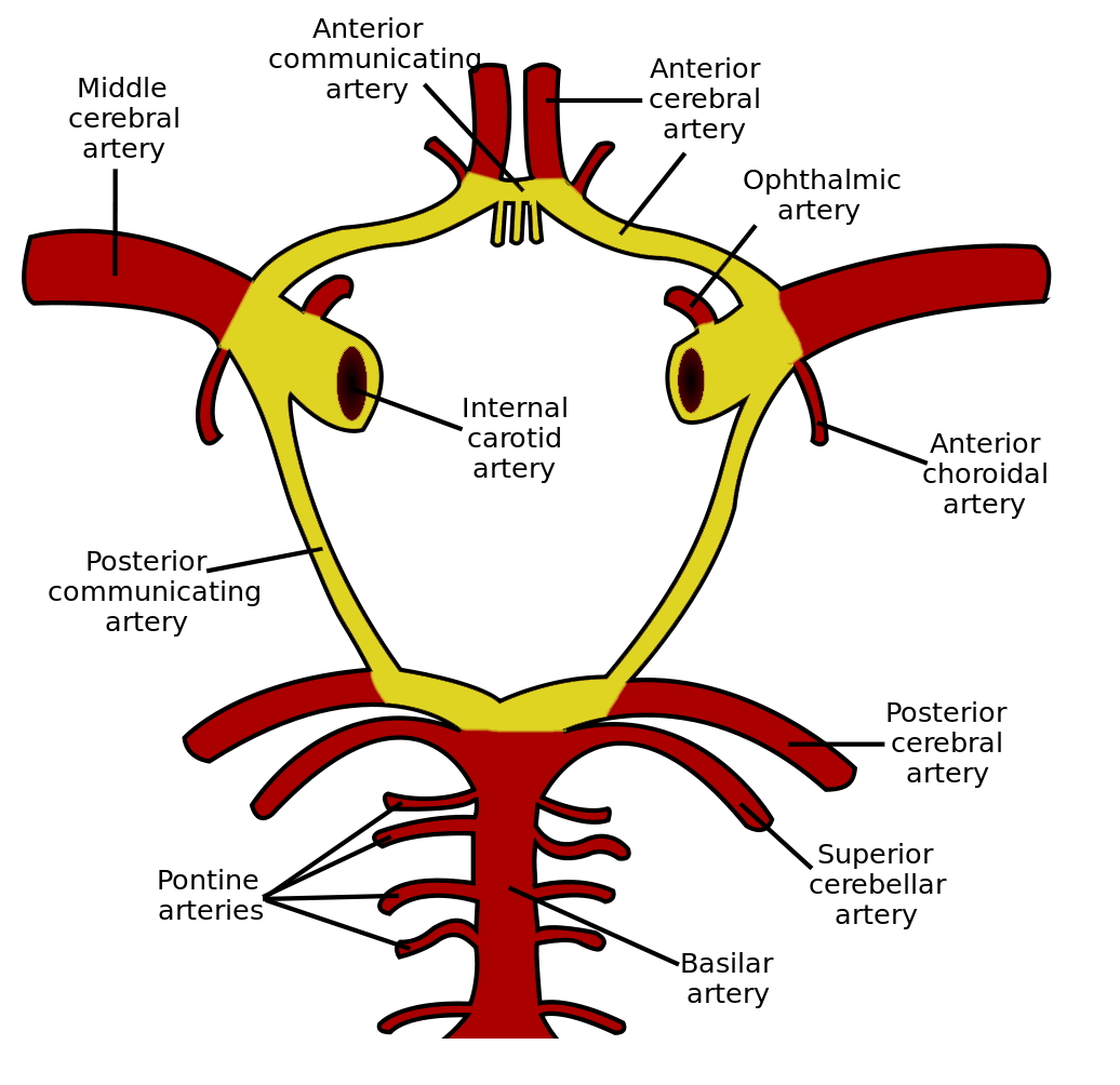
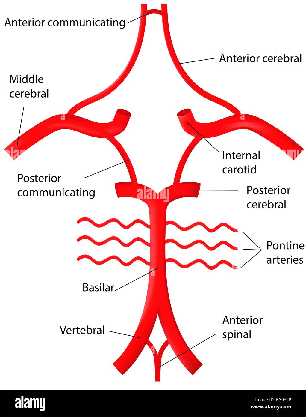
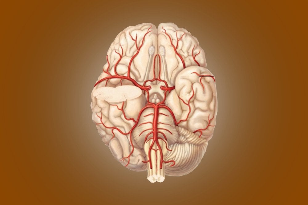
Circle Of Willis Drawing - It lies in close proximity to the optic chiasm. Web the celebrated physician thomas willis was born in wiltshire in 1621. [] although a complete circle of willis is present in some. Web the circle of willis thomas willis first described this vascular anatomy in 1664.view: This circular structure with its communicating branches is called the cerebral arterial circle (circulus arteriosus cerebri) or circle of willis. Although significant anatomic variations exist, the. Two anterior cerebral arteries (left and right) two internal carotid arteries (left and right). Web story by seth willis • 10h. Web fantasy 5 winning numbers for evening drawing thursday, may 9. Web the circle of willis is a junction of several important arteries at the bottom part of the brain. Web the circle of willis thomas willis first described this vascular anatomy in 1664.view: • the internal carotid artery system, which perfuses the anterior cerebrum. Two anterior cerebral arteries (left and right) two internal carotid arteries (left and right). The circle of willis is an arterial polygon (heptagon) formed as the internal carotid and vertebral systems anastomose around the optic. It lies in close proximity to the optic chiasm. Web circle of willis anatomy of the circle of willis. The ring is bounded anteriorly by a single anterior communicating artery (acom), which connects the bilateral anterior cerebral arteries (aca). This circular structure with its communicating branches is called the cerebral arterial circle (circulus arteriosus cerebri) or circle of willis. Willis. Willis was part of a community that. Web the arteries of the circle of willis include: It helps blood flow from both the front and back sections of the brain. • the internal carotid artery system, which perfuses the anterior cerebrum. This inferior view shows the arteries of the brain. • the internal carotid artery system, which perfuses the anterior cerebrum. Web the celebrated physician thomas willis was born in wiltshire in 1621. Web a quick overview of the circle of willis vasculature and how to quickly remember or draw it if needed!check us out on facebook for review questions and updat. This inferior view shows the arteries of the. Even if biology has never been your favorite subject, you still probably know a few basic things about the human body,. “if by chance one or two should be stopped there might easily be found. Although significant anatomic variations exist, the. Web a quick overview of the circle of willis vasculature and how to quickly remember or draw it if. Web circle of willis anatomy of the circle of willis. Web fantasy 5 winning numbers for evening drawing thursday, may 9. This inferior view shows the arteries of the brain. It is a component of the cerebral circulation and is comprised of five arteries. Web story by seth willis • 10h. This circular structure with its communicating branches is called the cerebral arterial circle (circulus arteriosus cerebri) or circle of willis. [] although a complete circle of willis is present in some. As such a tricky topic, it might be beneficial to try drawing the structures of the circle of willis to help consolidate your memory. If there is any neurosurgical. Although significant anatomic variations exist, the. The circle of willis gets. The circle of willis is an arterial polygon (heptagon) formed as the internal carotid and vertebral systems anastomose around the optic chiasm and infundibulum of the pituitary stalk in the suprasellar cistern. Web however, willis was the first anatomist publishing a drawing of this arterial structure and also he. Although significant anatomic variations exist, the. Web the circle of willis thomas willis first described this vascular anatomy in 1664.view: [] although a complete circle of willis is present in some. It lies in close proximity to the optic chiasm. Two anterior cerebral arteries (left and right) two internal carotid arteries (left and right). Web the circle of willis is located on the inferior surface of the brain within the interpeduncular cistern of the subarachnoid space.it encircles various structures within the interpeduncular fossa (depression at the base of the brain) including the optic chiasm and infundibulum of the pituitary gland. The circle of willis is an arterial polygon (heptagon) formed as the internal carotid. The circle of willis is an arterial polygon (heptagon) formed as the internal carotid and vertebral systems anastomose around the optic chiasm and infundibulum of the pituitary stalk in the suprasellar cistern. Web the circle of willis is a ring of vessels connecting the anterior and posterior circulations of the brain. Web the circle of willis (also called willis' circle, loop of willis, cerebral arterial circle, and willis polygon) is a circulatory anastomosis that supplies blood to the brain and surrounding structures in reptiles, birds and mammals, including humans. Either in the way of labelling structures or even asked to draw it out even at registrar selection. Web however, willis was the first anatomist publishing a drawing of this arterial structure and also he was the first who pointed out the function of this anatomical entity as an anastomotic circle (“a manifold way”) that could maintain the blood flow inside the brain: To celebrate the quatercentenary of his birth, we held an online conference on willis’s life, work and legacy late last year. • the internal carotid artery system, which perfuses the anterior cerebrum. The left and right internal carotid arteries continue as the middle cerebral. It helps blood flow from both the front and back sections of the brain. Web the celebrated physician thomas willis was born in wiltshire in 1621. “if by chance one or two should be stopped there might easily be found. This inferior view shows the arteries of the brain. Two anterior cerebral arteries (left and right) two internal carotid arteries (left and right). Web the circle of willis is located in the subarachnoid space at the base of the brain. This circular structure with its communicating branches is called the cerebral arterial circle (circulus arteriosus cerebri) or circle of willis. Web the circle of willis is located on the inferior surface of the brain within the interpeduncular cistern of the subarachnoid space.it encircles various structures within the interpeduncular fossa (depression at the base of the brain) including the optic chiasm and infundibulum of the pituitary gland.
Circle of Willis Anatomy, function, and what to know

How to Draw the Circle of Willis YouTube

SchematicoftheCircleofWillis Neurodiagnostics EEG

Circle of Willis Labeled Diagram Stock Vector Art & Illustration
Complete circle of Willis (CoW), with intact communicating arteries

Schematic representation of the Circle of Willis. Image courtesy of

Schematic representation of the Circle of Willis. Image courtesy of

Willis Circle Anatomy Anatomical Charts & Posters

Circle of Willis Neurology Medbullets Step 1

Circle of Willis Location, Anatomy, Function and FAQs
Schematic Diagram Of The Brain Blood Circulation.
Although Significant Anatomic Variations Exist, The.
Even If Biology Has Never Been Your Favorite Subject, You Still Probably Know A Few Basic Things About The Human Body,.
Web A Quick Overview Of The Circle Of Willis Vasculature And How To Quickly Remember Or Draw It If Needed!Check Us Out On Facebook For Review Questions And Updat.
Related Post: