Endometrial Polyp Size Chart In Mm
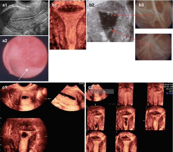
Endometrial Polyp Size Chart In Mm - Endometrial polyps form from an overgrowth of cells within the uterine lining. Polypectomy was done and the same was sent for histopathological evaluation. They may be a cause of menorrhagia and of post menopausal bleeding. Web an endometrial polyp represents the extreme end of macroscopic hyperplasia of the endometrium when tissue grows so fast that parts of the endometrium are pushed into the cavity of the uterus. Web a hysteroscopy is an examination procedure that is used to look inside the cavity of the uterus (womb) and see the polyps that are to be removed. Removal of asymptomatic polyps in premenopausal women should be considered in patients with risk factors for endometrial cancer (level b). Web the sonographic finding suggestive of an endometrial polyp is a bright, hyperechoic area visualized within the endometrium (table 1). Polyps may be found as a single lesion or multiple lesions filling the entire endometrial cavity. Web endometrial polyps greater than 15mm showed a hyperplasia rate of 14.8%, compared with 7.7% in the group with smaller polyps (p<0.05). Incidence in asymptomatic females with infertility: Endometrial polyps are localized tumors within the mucosa of the uterine cavity. Web asymptomatic endometrial polyps in postmenopausal women should be removed in case of large diameter (> 2 cm) or in patients with risk factors for endometrial carcinoma (level b). On hysteroscopy, there was hyperplastic endometrium with large endometrial polyp of size 8.5 cm. Can range in size from. They may be a cause of menorrhagia and of post menopausal bleeding. Polypectomy was done and the same was sent for histopathological evaluation. Polyps may be found as a single lesion or multiple lesions filling the entire endometrial cavity. Web the sonographic finding suggestive of an endometrial polyp is a bright, hyperechoic area visualized within the endometrium (table 1). Abnormal. However, peak incidence occurs between the age of 40 to 49 years old. Removal of asymptomatic polyps in premenopausal women should be considered in patients with risk factors for endometrial cancer (level b). Can range in size from millimeters (about the size of a sesame seed) to centimeters (about the size of a golf ball and even larger). Endometrial polyps. Endometrial polyps form from an overgrowth of cells within the uterine lining. Incidence in asymptomatic females with infertility: Can affect up to 25% of females presenting with abnormal uterine bleeding ( case rep obstet gynecol 2014;2014:518398 ) prevalence in asymptomatic females: Polyps may be round or oval and range in size from a few millimeters (the size of a sesame. Endometrial polyps may be diagnosed at all ages; Web an endometrial polyp or uterine polyp is a mass in the inner lining of the uterus. They may be a cause of menorrhagia and of post menopausal bleeding. Web the mean polyp size was 17.7 ± 0.5 mm in benign patients and 23.7 ± 1.8 mm in premalignant/malignant individuals ( p. In women below the age of 30 years, the prevalence was 0.9%. On hysteroscopy, there was hyperplastic endometrium with large endometrial polyp of size 8.5 cm. Web asymptomatic endometrial polyps in postmenopausal women should be removed in case of large diameter (> 2 cm) or in patients with risk factors for endometrial carcinoma (level b). Endometrial polyps refer to overgrowths. On average, these polyps are typically less than 1 cm. They may have a large flat base ( sessile) or be attached to the uterus by an elongated pedicle ( pedunculated ). In women below the age of 30 years, the prevalence was 0.9%. Endometrial polyps are common findings, both in women with and without gynaecological symptoms. Can range in. Endometrial polyps measuring more than 15mm were associated with hyperplasia. Web an endometrial polyp or uterine polyp is a mass in the inner lining of the uterus. [2] [3] pedunculated polyps are more common than sessile ones. Web the prevalence of endometrial polyps was 7.8% (48/619; A mostly benign pathology finding. Endometrial polyps are common findings, both in women with and without gynaecological symptoms. Can affect up to 25% of females presenting with abnormal uterine bleeding ( case rep obstet gynecol 2014;2014:518398 ) prevalence in asymptomatic females: They contain glands, connective tissues, and blood vessels. Removal of asymptomatic polyps in premenopausal women should be considered in patients with risk factors for. Endometrial polyps are common findings, both in women with and without gynaecological symptoms. Endometrial polyps refer to overgrowths of endometrial glands and stroma within the uterine cavity. Can range in size from millimeters (about the size of a sesame seed) to centimeters (about the size of a golf ball and even larger). Web a hysteroscopy is an examination procedure that. Endometrial polyps measuring more than 15mm were associated with hyperplasia. The lesions may contain blood vessels and cause irregular menstrual bleeding, spotting, menorrhagia, and postmenopausal bleeding. Web an endometrial polyp or uterine polyp is an abnormal growth containing glands, stroma and blood vessels projecting from the lining of the uterus (endometrium) that occupies spaces small or large enough to fill the uterine cavity. Web a hysteroscopy is an examination procedure that is used to look inside the cavity of the uterus (womb) and see the polyps that are to be removed. [2] [3] pedunculated polyps are more common than sessile ones. They also range in number women can have one or many endometrial polyps. Web the polyp attaches to the endometrium by a thin stalk or a broad base and extends into your uterus. They contain glands, connective tissues, and blood vessels. Web an endometrial polyp or uterine polyp is a mass in the inner lining of the uterus. Endometrial polyps form from an overgrowth of cells within the uterine lining. Polyps may be round or oval and range in size from a few millimeters (the size of a sesame seed) to a few centimeters (the size of a golf ball) or larger. Web patient was planned for hysteroscopic guided biopsy as her ultrasonography (usg) showed endometrial thickness to be 12.3 mm. Web asymptomatic endometrial polyps in postmenopausal women should be removed in case of large diameter (> 2 cm) or in patients with risk factors for endometrial carcinoma (level b). They may be a cause of menorrhagia and of post menopausal bleeding. Polyps may be found as a single lesion or multiple lesions filling the entire endometrial cavity. Endometrial polyps vary in size from a few millimeters to several centimeters in diameter.
Clinical by individual endometrial thickness Download Table
![[PDF] Giant endometrial polyp protruding from the external cervical os](https://d3i71xaburhd42.cloudfront.net/8ee776e2c239fe8f6fe5bef07581c99c4de87bae/5-Figure4-1.png)
[PDF] Giant endometrial polyp protruding from the external cervical os

Endometrial Polyp Size Chart

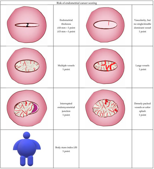
Diagnostics Free FullText Risk Assessment of Endometrial

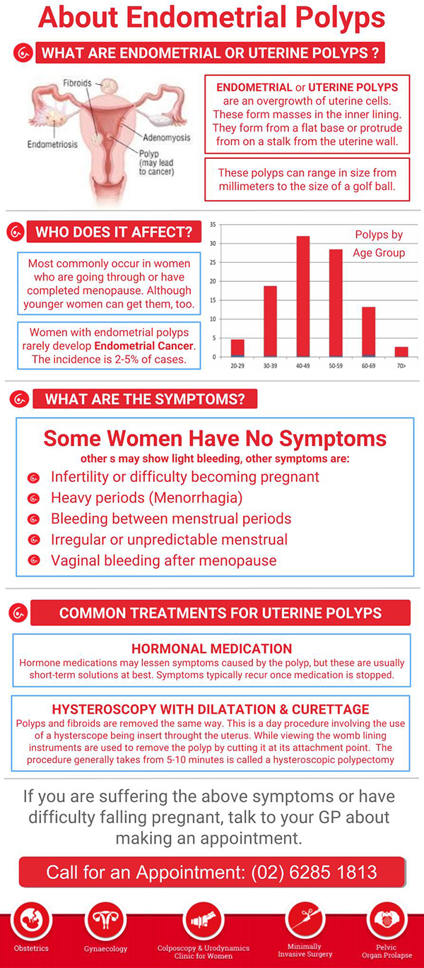
Endometrial & Uterine Polyps Canberra Deakin, ACT

Narrowband imaging without high magnification to differentiate polyps
Representative size measurement and appearance of endometrial polyps

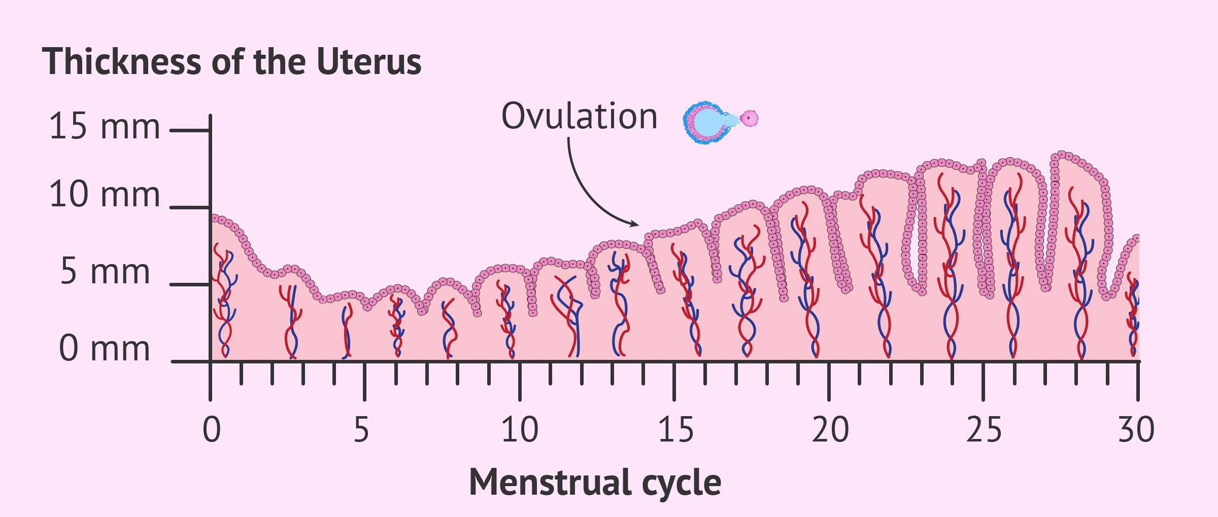
Endometrial Lining Thickness Chart Labb by AG

Endometrial Polyp Size Chart In Mm

Uterine Polyp Size Chart
Web Hysteroscopy With Guided Biopsy Remains The Gold Standard For Diagnosis Of Endometrial Polyps (High).
Overgrowth Of Localized Endometrial Tissue That May Be Pedunculated Or Sessile, Single Or Multiple, And Up To Many Centimeters In Size.
On Average, These Polyps Are Typically Less Than 1 Cm.
Endometrial Polyps Are Localized Tumors Within The Mucosa Of The Uterine Cavity.
Related Post: