Eukaryotic Cell Drawing
Eukaryotic Cell Drawing - The cell wall is a rigid covering that protects the cell, provides structural support, and gives shape to the cell. Made of α and β tubulin combined to form dimers, the dimers are then joined into protofilaments. If you examine figure 4.8, the plant cell diagram, you will see a structure external to the plasma membrane. These organisms are grouped into the biological domain eukaryota. For the purpose of this article, the primary focus will be the structure and histology of the animal cell. Web in eukaryotic cells, the membrane that surrounds the nucleus — commonly called the nuclear envelope — partitions this dna from the cell's protein synthesis machinery,. Ribosomes and lyosomes and a number of tiny filament. The cell membrane is represented as the factory walls. the nucleus of a cell is represented as the blueprint room.. Actin is both flexible and strong, making it a useful protein in cell movement. Fungal and some protistan cells also have cell walls. Made of α and β tubulin combined to form dimers, the dimers are then joined into protofilaments. This is the cell wall, a rigid covering that protects the cell, provides structural support, and gives shape to the cell. Prokaryotes are straightforward, using just one type of rna polymerase to transcribe their dna into rna. Ribosomes and lyosomes and a number. A diagram showing the basic structure of a eukaryotic cell and prokaryotic cell. The cell wall is a rigid covering that protects the cell, provides structural support, and gives shape to the cell. The cell cycle length is highly variable within the different cell types. Eukaryotic cells also contain organelles, including mitochondria (cellular energy exchangers), a. Web the cell wall. Web diagram and micrograph of intestinal cells, showing the protruding fingers of plasma membrane—called microvilli—that contact the fluid inside the small intestine. Web like prokaryotes, eukaryotic cells have a plasma membrane. In addition to the nucleus, eukaryotic cells. Thirteen protofilaments in a cylinder make a microtubule. Organisms based on the eukaryotic cell include protozoa, fungi, plants, and animals. In figure 1b, the diagram of a plant cell, you see a structure external to the plasma membrane called the cell wall. For epithelial cells in humans, it is about two to five days. Web a diagram representing the cell as a factory. Web drawing eukaryotic cells and annotating the functions of each of the organelles. This is the cell. Made of α and β tubulin combined to form dimers, the dimers are then joined into protofilaments. They're also the more complex of the two. Structure and function is shared under a not declared license and was authored, remixed, and/or curated by libretexts. Web a eukaryotic cell cycle is an ordered event involving cell growth and division, producing two daughter. Flagella and cilia are the locomotory organs in a eukaryotic cell. Web in eukaryotic cells, the membrane that surrounds the nucleus — commonly called the nuclear envelope — partitions this dna from the cell's protein synthesis machinery,. Makes up the cytoskeleton of the cell about 25 nm in diameter. For epithelial cells in humans, it is about two to five. The major differences between animal and plant cells will be explored as well. As previously stated, the fundamental components of a cell are its organelles. Prokaryotes are straightforward, using just one type of rna polymerase to transcribe their dna into rna. Actin is both flexible and strong, making it a useful protein in cell movement. Web a diagram representing the. Please note, that usually the cells are so densely packed with structures, that if this was an accurate representation of the amount of components, it would be. Thirteen protofilaments in a cylinder make a microtubule. In prokaryotes, which lack a nucleus, cytoplasm. Eukaryotic cells are larger and more complex than. Structure and function is shared under a not declared license. In addition to the nucleus, eukaryotic cells. In figure 1b, the diagram of a plant cell, you see a structure external to the plasma membrane called the cell wall. Web characteristics of eukaryotic cells. Web about press copyright contact us creators advertise developers terms privacy policy & safety how youtube works test new features nfl sunday ticket press copyright. An. An early embryonic cell has a turnover range of a few hours. The cell wall is a rigid covering that protects the cell, provides structural support, and gives shape to the cell. Eukaryotes, however, use three different rna polymerases, allowing them a more tailored approach to transcribing dna. The cell membrane is represented as the factory walls. the nucleus of. Web characteristics of eukaryotic cells. Flagella and cilia are the locomotory organs in a eukaryotic cell. Web in eukaryotic cells, the membrane that surrounds the nucleus — commonly called the nuclear envelope — partitions this dna from the cell's protein synthesis machinery,. A plasma membrane encloses the cell contents of both plant and animal cells, but it is the outer coating of an animal cell. They're also the more complex of the two. Please note, that usually the cells are so densely packed with structures, that if this was an accurate representation of the amount of components, it would be. Eukaryotic cells have the nucleus enclosed within the nuclear membrane. A 3d model of a eukaryote including the major components, while missing a few smaller structures: Organisms based on the eukaryotic cell include protozoa, fungi, plants, and animals. For epithelial cells in humans, it is about two to five days. These organisms are grouped into the biological domain eukaryota. Fungal and some protistan cells also have cell walls. Fungal and protist cells also have cell walls. As previously stated, the fundamental components of a cell are its organelles. In prokaryotes, which lack a nucleus, cytoplasm. The cell cycle length is highly variable within the different cell types.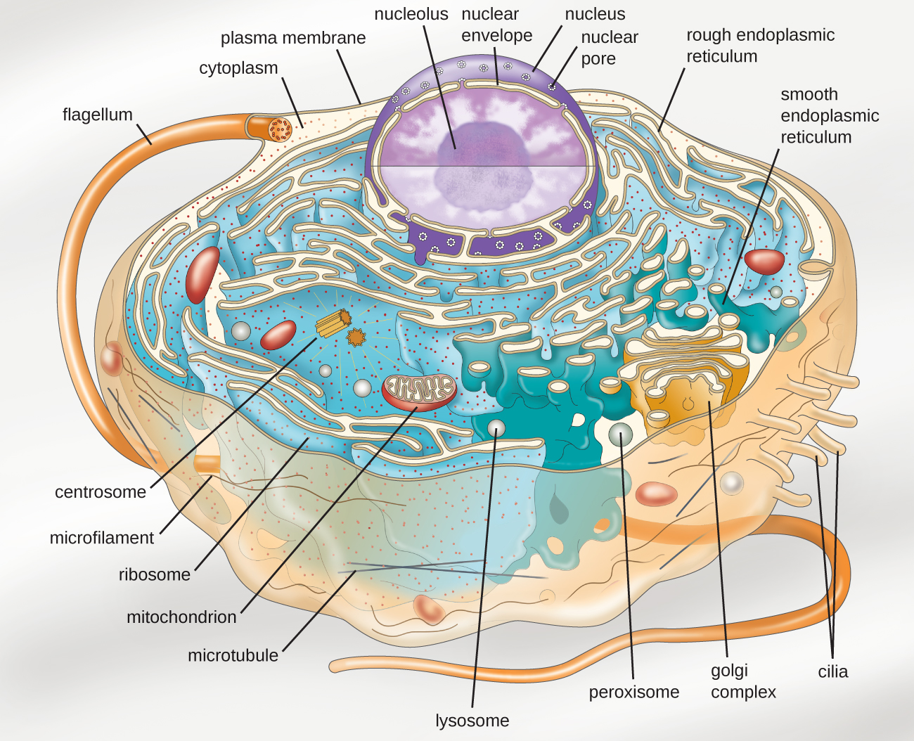
3.4 Unique Characteristics of Eukaryotic Cells Microbiology 201

Eukaryotic Cell Diagram 2d
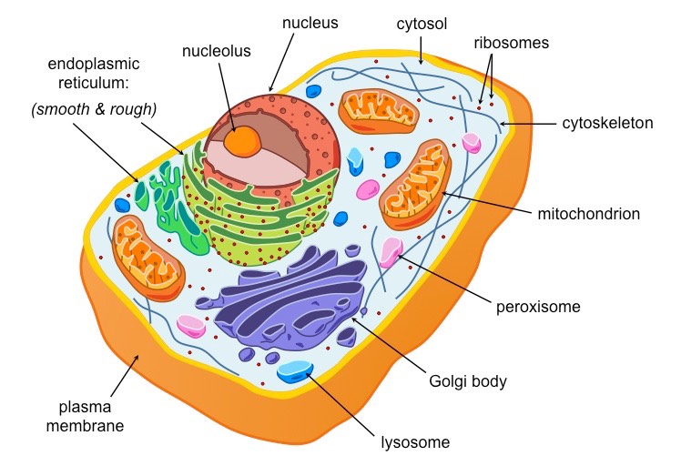
Characteristics Of Eukaryotic Cellular Structures ALevel Biology
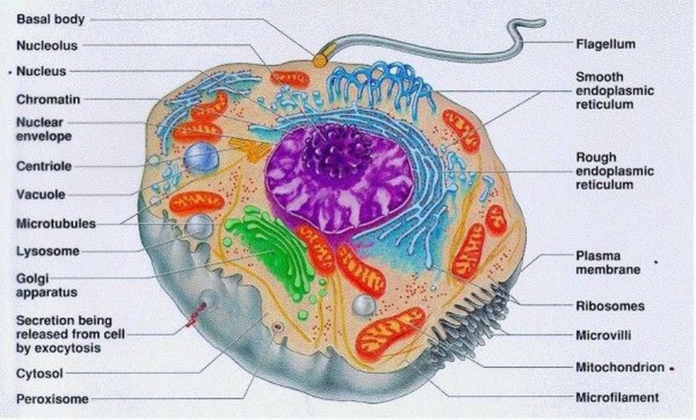
2.4 Eukaryotic Cell Structure a level biology student
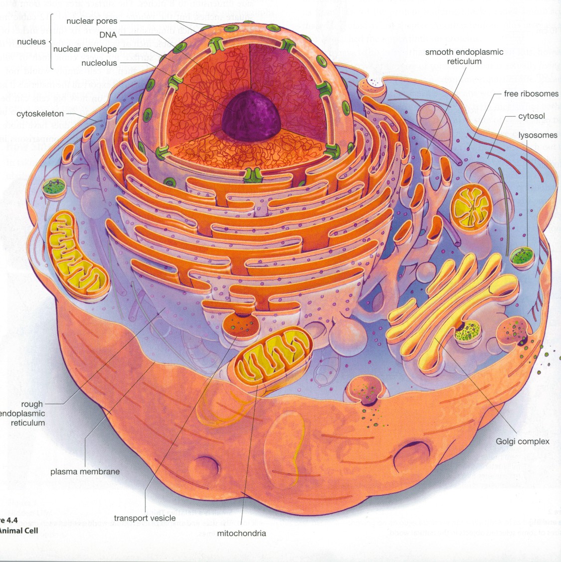
Eukaryotic cell structure diagrams Biological Science Picture

Eukaryotic Cell Diagram 2d

How to Draw an Animal Cell Really Easy Drawing Tutorial

Biology 2e, The Cell, Cell Structure, Eukaryotic Cells INFOhio Open Space
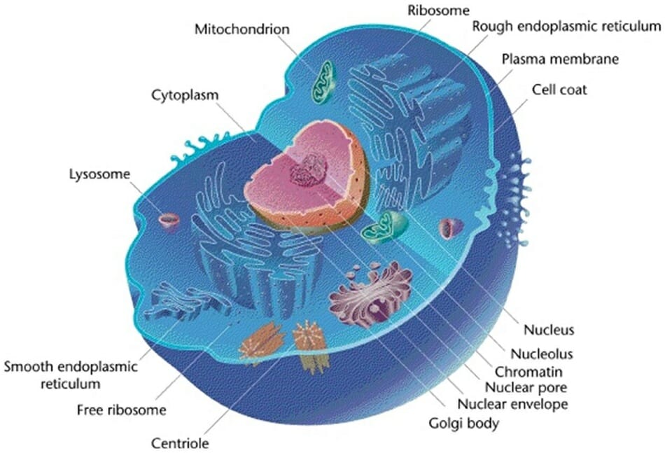
Eukaryotic Cell Definition, Characteristics, Structure and Examples

Parts of the Eukaryotic Cell Diagram Quizlet
Web Diagram And Micrograph Of Intestinal Cells, Showing The Protruding Fingers Of Plasma Membrane—Called Microvilli—That Contact The Fluid Inside The Small Intestine.
The Location Of The Dna Is Highlighted In Each.
Eukaryotes, However, Use Three Different Rna Polymerases, Allowing Them A More Tailored Approach To Transcribing Dna.
Web Figure 1 Below Shows A Simple Diagram Of Each.
Related Post: