Flow Chart Of A Cell
Flow Chart Of A Cell - All symbols in the flowchart must be connected with an arrow line. There are two methods for obtaining cells: To divide, a cell must complete several important tasks: Web a flow cytometer has five main components: Interphase is the time during which the cell prepares for division by undergoing both cell growth and dna replication. Organelles in animal cells include the nucleus, mitochondria, endoplasmic reticulum, golgi apparatus, vesicles, and vacuoles. Web flow cytometry is a widely used method for analyzing the expression of cell surface and intracellular molecules, characterizing and defining different cell types in a heterogeneous cell population, assessing the purity of isolated subpopulations, and analyzing cell size and volume. Web a data flow diagram (dfd) helps you understand how data flows through a system. Web a flowchart is a graphical representations of steps. Virtually every cell, tissue, organ, and system in the body is impacted by the circulatory system. Protocols for preparing cells for flow cytometry 44. Web a data flow diagram (dfd) helps you understand how data flows through a system. Web among these 6,094 cells, 3,839 cells (63%) had at least one rare allele (figure 3k), and this fraction was comparable across the seven blood cell types in the skull (figure 3l). The ux design is a. Organelles in animal cells include the nucleus, mitochondria, endoplasmic reticulum, golgi apparatus, vesicles, and vacuoles. Also known as the resting phase of the cell cycle; From a cell bank or by isolating cells from donor tissue. Preparation of tissue culture cells stored in liquid nitrogen 44. Web a data flow diagram (dfd) helps you understand how data flows through a. Web for example, consider the idea that cells are the building blocks of life. Web a data flow diagram (dfd) helps you understand how data flows through a system. Web a flowchart is a graphical representations of steps. Flowchart opening statement must be ‘start’ keyword. Some textbooks list five, breaking prophase into an early phase (called prophase) and a late. Web purpose this study aims to compare treatment outcomes between neoadjuvant chemotherapy (nact) followed by surgery and concurrent chemoradiotherapy (ccrt) in patients with stage iib cervical squamous cell carcinoma (cscc). A flow cell, a measuring system, a detector, an amplification system, and a computer for analysis of the signals. Types of user flow charts. Flowchart of a generalized cell culture. Web flow cytometry is a widely used method for analyzing the expression of cell surface and intracellular molecules, characterizing and defining different cell types in a heterogeneous cell population, assessing the purity of isolated subpopulations, and analyzing cell size and volume. Ribosomes are not enclosed within a membrane but are still commonly referred to as organelles in eukaryotic cells. To. It occupies around 95% time of the overall cycle. Preparation of adherent tissue culture cell lines 45. Ribosomes are not enclosed within a membrane but are still commonly referred to as organelles in eukaryotic cells. Types of user flow charts. From a cell bank or by isolating cells from donor tissue. After completing the cycle it either starts the process again from g1 or exits through g0. Web a flow cytometer has five main components: Web the basic flow of genetic information in biological systems is often depicted in a scheme known as the central dogma (see figure below). Protocols for preparing cells for flow cytometry 44. Prophase, metaphase, anaphase, and. A common task of many research teams is the analysis of cell cycle progression through the distinct cell cycle phases. Web a flowchart is a graphical representation of an algorithm.it should follow some rules while creating a flowchart. To divide, a cell must complete several important tasks: Web a data flow diagram (dfd) helps you understand how data flows through. Ribosomes are not enclosed within a membrane but are still commonly referred to as organelles in eukaryotic cells. Preparation of adherent tissue culture cell lines 45. From a cell bank or by isolating cells from donor tissue. In our muscle cells, when there is lack of oxygen glucose breaks down to form ethanol, carbon dioxide and energy. If that idea. Various methods can be used for this purpose. A dfd simplifies complex processes by breaking them down into interconnected bubbles and arrows, creating a visual representation of how data moves and changes. When starting culture from cells obtained from a cell bank, one needs to go through the procedures of thawing, cell seeding and cell observation. Preparation of tissue culture. Web one of the fundamentals of flow cytometry is the ability to measure the properties of individual particles. By using standardized symbols and definitions, you can create a handy visual representation of any process's various steps and decision points. Organelles in animal cells include the nucleus, mitochondria, endoplasmic reticulum, golgi apparatus, vesicles, and vacuoles. Ribosomes are not enclosed within a membrane but are still commonly referred to as organelles in eukaryotic cells. Web explain the process of breakdown of glucose in a cell. In fact, we do observe this (our actual observation), so evidence supports the idea that living things are built from cells. A flow cell, a measuring system, a detector, an amplification system, and a computer for analysis of the signals. The flow cell has a liquid stream (sheath fluid), which carries and aligns the cells so that they pass single file through the light beam for sensing. Prophase, metaphase, anaphase, and telophase. To divide, a cell must complete several important tasks: Web organelles are involved in many vital cell functions. Web for example, consider the idea that cells are the building blocks of life. Web a flow cytometer has five main components: If that idea were true, we’d expect to see cells in all kinds of living tissues observed under a microscope — that’s our expected observation. From a cell bank or by isolating cells from donor tissue. (ii) in absence of oxygen.
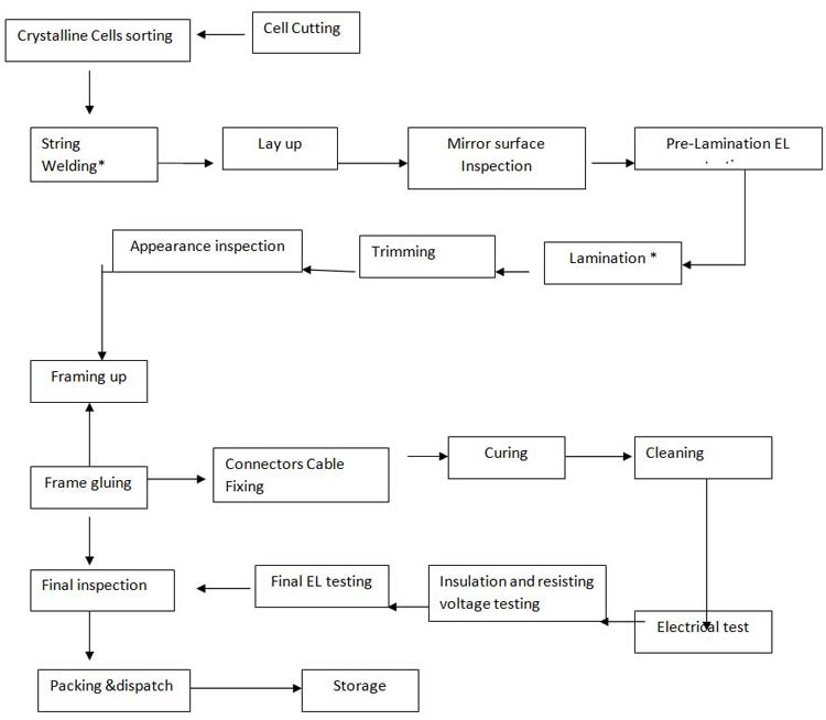
The flow chart illustrates collection, processing and storage of

Parts Of A Cell Flow Chart Kemele

What Stimulates Cell Division Brainly Cell cycle presentation by

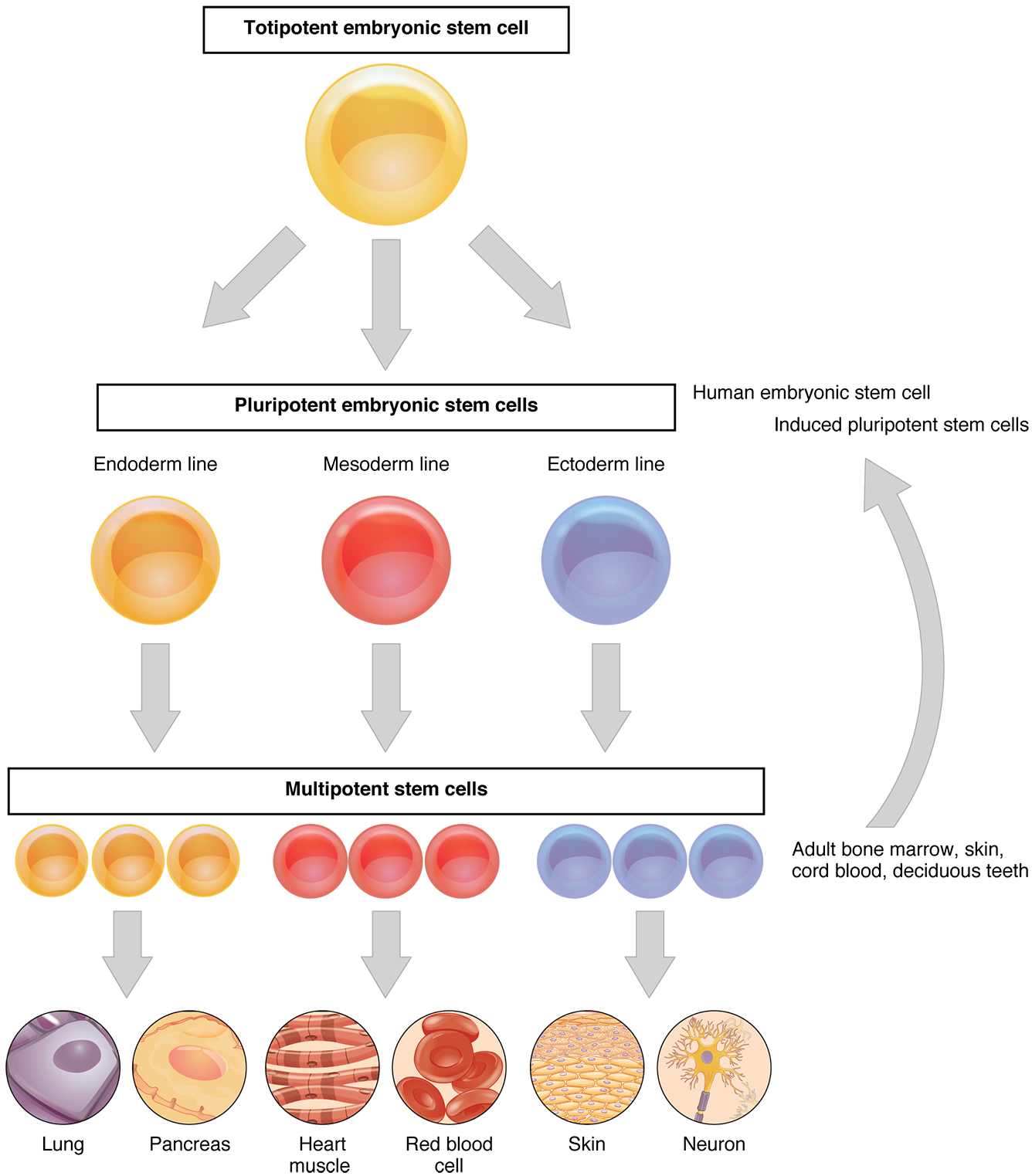
Cellular Differentiation Anatomy and Physiology I

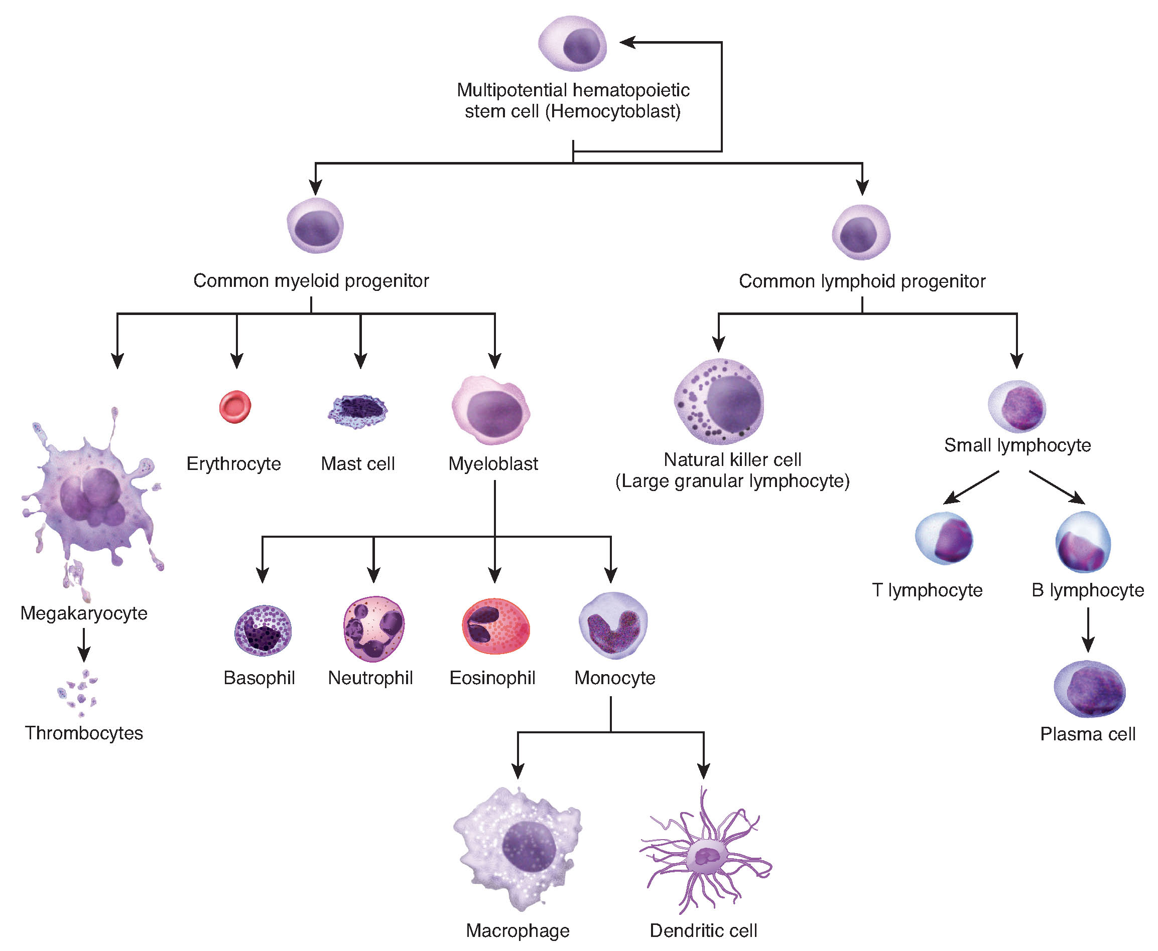
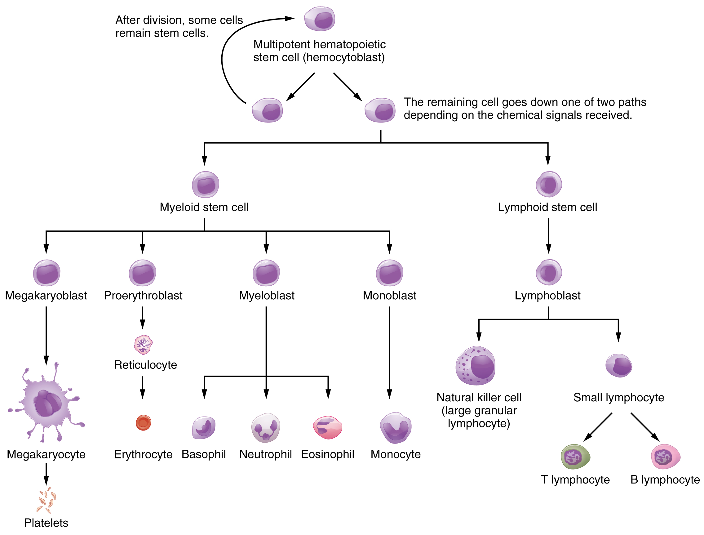
This flowchart shows the differentiation of a hemocytoblast, a stem

Cell Division Flowchart Cell Division

Parts Of A Cell Flow Chart Kemele

cell and structure flowchart Brainly.in

Production of the Formed Elements · Anatomy and Physiology

This flow chart shows the differentiation of stem cells into different
A Common Task Of Many Research Teams Is The Analysis Of Cell Cycle Progression Through The Distinct Cell Cycle Phases.
Web Create A Flow Chart Showing The Major Systemic Veins Through Which Blood Travels From The Feet To The Right Atrium Of The Heart.
There Are Two Methods For Obtaining Cells:
Web A Flowchart Is A Graphical Representations Of Steps.
Related Post: