H Wave Electrode Placement Chart
H Wave Electrode Placement Chart - Bolus manipulation and tongue base retraction. A visual representation of the cervical thoracic electrode placement. First electrode is placed well above hyoid bone. These videos are only examples to give a sense of how h‑wave is applied. 65% of respondents reported a reduced need for medication, 79% reported improved functionality, and 78% reported a 25% or greater. Clean your skin with a damp washcloth in the location the pads are placed. Attach pads per the illustration on the right for pain on the inside of your ankle/foot. Web a visual representation of a typical lumbar and sacral electrode placement for pain and low back issues. Ankle/foot attach pads per the illustration on the left for pain on the outside of your ankle/foot. & “high” for b (65 pps) raise amplitude for b until a strong tingling sensation, less than muscle contraction. Clean your skin with a damp washcloth in the location the pads are placed. Place pads at least 1 inch apart on clean, dry, healthy skin. All electrodes aligned vertically along midline. 1 the v1 electrode should be placed to the right of the sternal border, and v2 should be placed at the left of the sternal border in. A. Web pads are placed in pairs, and therapy will be felt between each pair of electrodes. Web the videos below show examples of electrode setups and how your body can be positioned during treatment. 65% of respondents reported a reduced need for medication, 79% reported improved functionality, and 78% reported a 25% or greater. A visual representation of a typical. A visual representation of the cervical thoracic electrode placement. 65% of respondents reported a reduced need for medication, 79% reported improved functionality, and 78% reported a 25% or greater. Web place electrode pads on both legs at the same time). Functional muscle actions possible signs & symptoms possible vitalstim electrode placements oropharyngeal “sling”. Bottom electrode should not end up below. Web place electrode pads on both legs at the same time). Find out the features, operation, pad placement, and contraindications of the device. & “high” for b (65 pps) raise amplitude for b until a strong tingling sensation, less than muscle contraction. 65% of respondents reported a reduced need for medication, 79% reported improved functionality, and 78% reported a 25%. Apply pain relief pads directly over areas of pain. A visual representation of the cervical thoracic electrode placement. Web cervical thoracic placement example. Actual instruction developed by your h‑wave consultant and doctor is customized for you and can vary significantly. All electrodes aligned vertically along midline. Web place electrode pads on both legs at the same time). & “high” for b (65 pps) raise amplitude for b until a strong tingling sensation, less than muscle contraction. Select “low” for channel a (1.5 pps); 3rd and 4th electrode placed at equal distances below first two electrodes. Actual instruction developed by your h‑wave consultant and doctor is customized. 1 the v1 electrode should be placed to the right of the sternal border, and v2 should be placed at the left of the sternal border in. All electrodes aligned vertically along midline. Find out the features, operation, pad placement, and contraindications of the device. It has become a staple in our core service provisions and is one of the. Attach pads per the illustration on the right for pain on the inside of your ankle/foot. Actual instruction developed by your h‑wave consultant and doctor is customized for you and can vary significantly. 3rd and 4th electrode placed at equal distances below first two electrodes. Find out the features, operation, pad placement, and contraindications of the device. Clean your skin. Web a visual representation of a typical lumbar and sacral electrode placement for pain and low back issues. 3rd and 4th electrode placed at equal distances below first two electrodes. Web pads are placed in pairs, and therapy will be felt between each pair of electrodes. Functional muscle actions possible signs & symptoms possible vitalstim electrode placements oropharyngeal “sling”. Web. Select “low” for channel a (1.5 pps); 65% of respondents reported a reduced need for medication, 79% reported improved functionality, and 78% reported a 25% or greater. Attach pads per the illustration on the right for pain on the inside of your ankle/foot. First electrode is placed well above hyoid bone. Patients will be instructed on optimal electrode placement for. All electrodes aligned vertically along midline. Bolus manipulation and tongue base retraction. Second electrode is placed just below first one, above the thyroid notch. Web the videos below show examples of electrode setups and how your body can be positioned during treatment. 1 the v1 electrode should be placed to the right of the sternal border, and v2 should be placed at the left of the sternal border in. 12k views 5 years ago. Apply pain relief pads directly over areas of pain. Actual instruction developed by your h‑wave consultant and doctor is customized for you and can vary significantly. A visual representation of the cervical thoracic electrode placement. It has become a staple in our core service provisions and is one of the few devices that all our clinics are encouraged to. 3rd and 4th electrode placed at equal distances below first two electrodes. 65% of respondents reported a reduced need for medication, 79% reported improved functionality, and 78% reported a 25% or greater. Web pads are placed in pairs, and therapy will be felt between each pair of electrodes. & “high” for b (65 pps) raise amplitude for b until a strong tingling sensation, less than muscle contraction. Patients will be instructed on optimal electrode placement for home use. Select “low” for channel a (1.5 pps);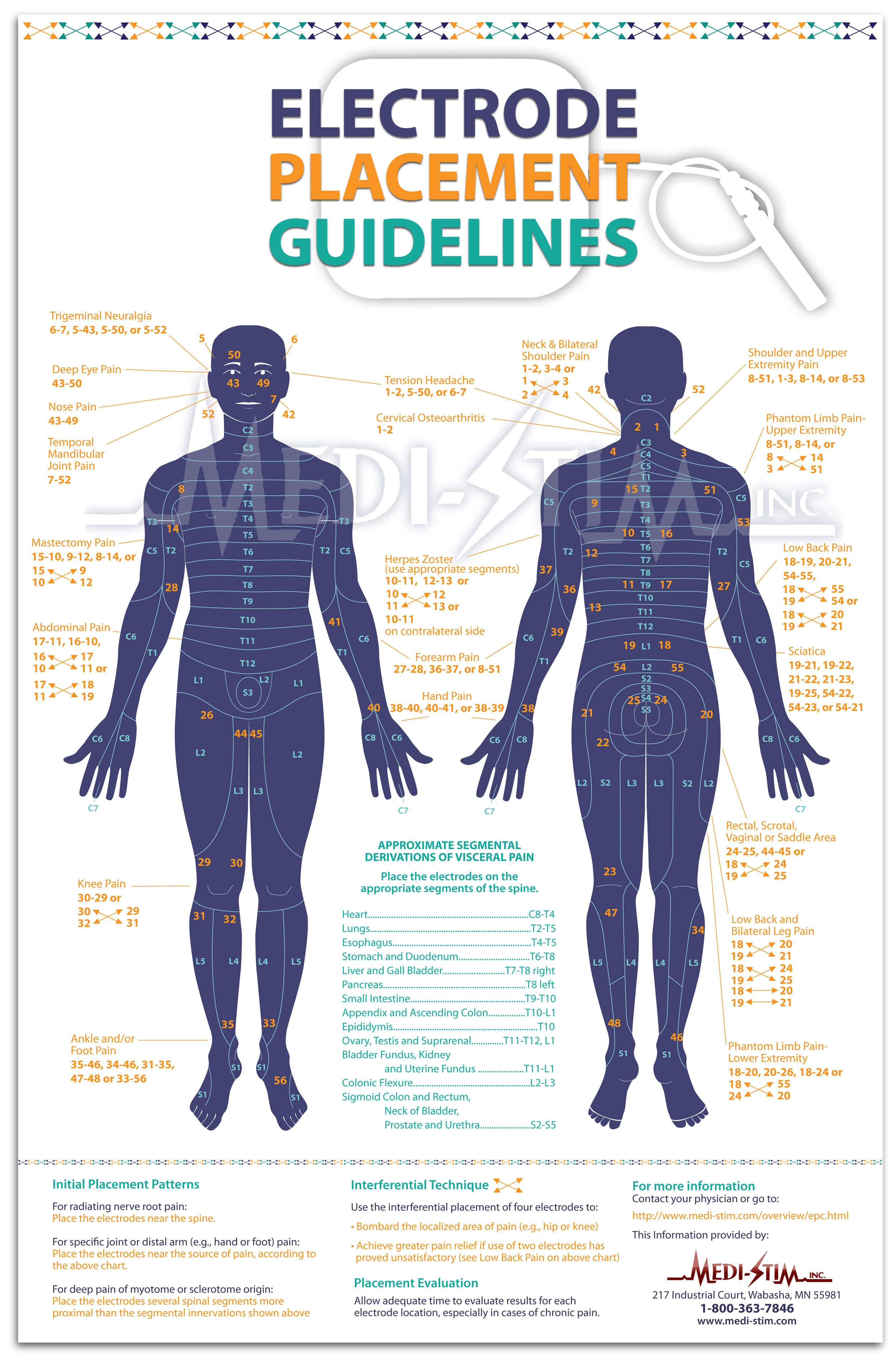
Electrode Placement Chart TENS Electrode Guidelines MediStim, Inc.

HWave YouTube

Hwave Electrode Placement Chart
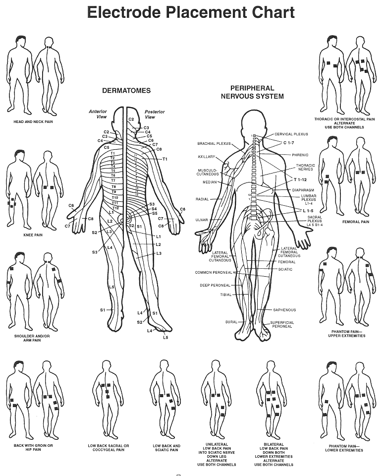
Electronic Pulse Massager
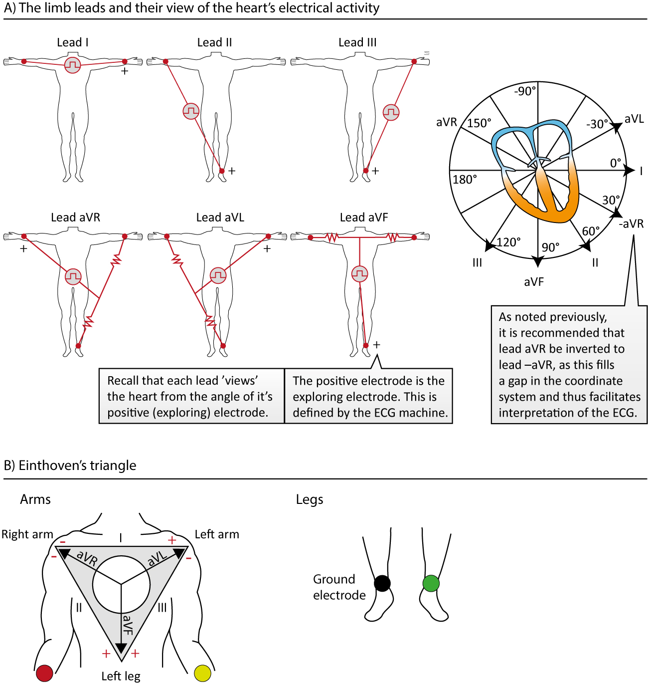
The ECG leads electrodes, limb leads, chest (precordial) leads, 12

H Wave Electrode Placement Chart
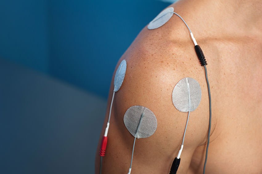
ShoulderPadPlacement HWave
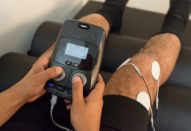
rightkneemale HWave

How To Do An Emg

TIBIAL HREFLEX YouTube
Ankle/Foot Attach Pads Per The Illustration On The Left For Pain On The Outside Of Your Ankle/Foot.
Web Connect Lead To The Electrodes An Place Electrodes On Skin According To Diagram (Ignore C Placement) Connect Leads To Channels Of H4 Device & Power On.
Functional Muscle Actions Possible Signs & Symptoms Possible Vitalstim Electrode Placements Oropharyngeal “Sling”.
Attach Pads Per The Illustration On The Right For Pain On The Inside Of Your Ankle/Foot.
Related Post: