Human Kvp And Mas Technique Chart
Human Kvp And Mas Technique Chart - Kvp and the emission spectra; The charts ordinarily work well and can be depended upon most of the time. Yes (ensure the correct grid is selected if using focused grids) image technical evaluation. Explain the effect on ir exposure, contrast, spatial resolution and distortion with either an increase or decrease in the following factors: 110 cm source image distance unless otherwise stated: Web general rule of thumb for ma settings for patient weight is: Description of the system(s) log 10 factor is the value needed to increase the parameter per cm. Web 35 cm x 43 cm. The entire lumbar spine should be visible, with demonstration of t11/t12 superiorly and the sacrum inferiorly. Web tube current (ma)* new tube current (ma)* tube potential (kvp)* new potential (kvp)* sid (mm)* new sid (mm)* bucky* new bucky* table of contents. Web log 10 technique chart: Web tube current (ma)* new tube current (ma)* tube potential (kvp)* new potential (kvp)* sid (mm)* new sid (mm)* bucky* new bucky* table of contents. You can use it to image animals in a veterinary setting and humans in a medical setting. Web general rule of thumb for ma settings for patient weight is: With. Web with a fixed kvp technique chart an optimal kvp is determined for each body part based upon contrast and penetrating quality; Web exposures shown in kvp/mas format. In smaller patients, the lower spectrum of the kv range is used; Determine 2 the starting peak potential (kvp) by taking a lateral abdominal film of. Kilovoltage, rectification and phase generation; Ma (tube current) kvp (tube current) s (time) sid (source to image distance) bucky factor (scatter grid factor) The 60 to 90 kvp range; Determine 2 the starting peak potential (kvp) by taking a lateral abdominal film of. The kvp must be accurate: 0.1 mm copper + 1 mm aluminium added beam filtration. Web exposures shown in kvp/mas format. 0.1 mm copper + 1 mm aluminium additional beam filtration. The kvp must be accurate: Explain the effect on ir exposure, contrast, spatial resolution and distortion with either an increase or decrease in the following factors: A fixed kvp technique chart is commonly used with automatic exposure control (aec) units. Kilovoltage, rectification and phase generation; G, grid exposure 8:1 ratio. This concept and procedure will be developed future later in the paper. But a good radiographer also has the ability to use different settings from those the technique chart calls for depending on variables that can occur in clinical practice. You can use it to image animals in a veterinary. Web the primary exposure technique factors the radiographer selects on the control panel are milliamperage (ma), time of exposure, and kilovoltage peak (kvp). Web the suggested technique is within a fixed kilovolt (kv) range per body part. In larger patients, the upper range of kv is used. The radiographer is responsible for selecting exposure factor techniques to produce quality radiographs. The radiographer is responsible for selecting exposure factor techniques to produce quality radiographs for a wide variety of equipment and patients. You can use it to image animals in a veterinary setting and humans in a medical setting. The kvp must be accurate: Web general rule of thumb for ma settings for patient weight is: Ma (tube current) kvp (tube. Yes (ensure the correct grid is selected if using focused grids) image technical evaluation. Ma (tube current) kvp (tube current) s (time) sid (source to image distance) bucky factor (scatter grid factor) G, grid exposure 8:1 ratio. Keep in mind when making your choice for the standard kvp that higher kvp will allow you to reduce the mas and reduce. A fixed kvp technique chart is commonly used with automatic exposure control (aec) units. In larger patients, the upper range of kv is used. Web variable kvp/fixed mas technique chart. The exposure must be linear: The radiographer is responsible for selecting exposure factor techniques to produce quality radiographs for a wide variety of equipment and patients. Kilovoltage, rectification and phase generation; The 60 to 90 kvp range; This chart is vital in the medical imaging field. Web exposures shown in kvp/mas format. The exposure must be linear: Revised 8/24/21 kvp selection chart for adult chest ct height in feet, inches weight. Web the suggested technique is within a fixed kilovolt (kv) range per body part. Web the primary exposure technique factors the radiographer selects on the control panel are milliamperage (ma), time of exposure, and kilovoltage peak (kvp). Depending on the type of control panel, milliamperage and exposure time may be selected separately or combined as one factor, milliamperage/second (mas). In larger patients, the upper range of kv is used. Determine 2 the starting peak potential (kvp) by taking a lateral abdominal film of. But a good radiographer also has the ability to use different settings from those the technique chart calls for depending on variables that can occur in clinical practice. 110 cm source image distance unless otherwise stated: A fixed kvp technique chart is commonly used with automatic exposure control (aec) units. Web tube current (ma)* new tube current (ma)* tube potential (kvp)* new potential (kvp)* sid (mm)* new sid (mm)* bucky* new bucky* table of contents. G, grid exposure 8:1 ratio. In smaller patients, the lower spectrum of the kv range is used; The entire lumbar spine should be visible, with demonstration of t11/t12 superiorly and the sacrum inferiorly. Web with a fixed kvp technique chart an optimal kvp is determined for each body part based upon contrast and penetrating quality; 0.1 mm copper + 1 mm aluminium added beam filtration. This chart is vital in the medical imaging field.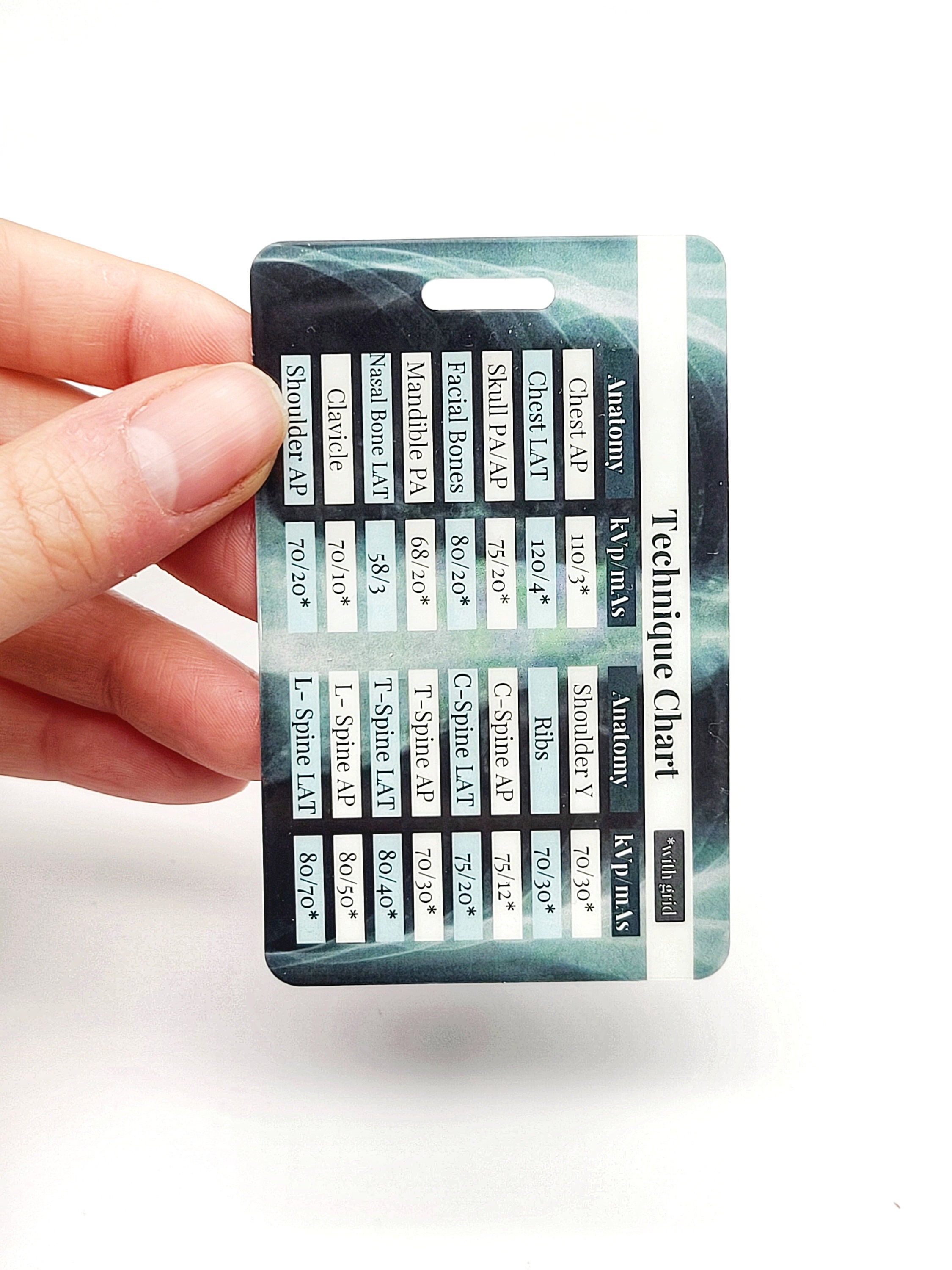
Xray Technique Card Xray Mas and Kvp Chart Radiology Etsy
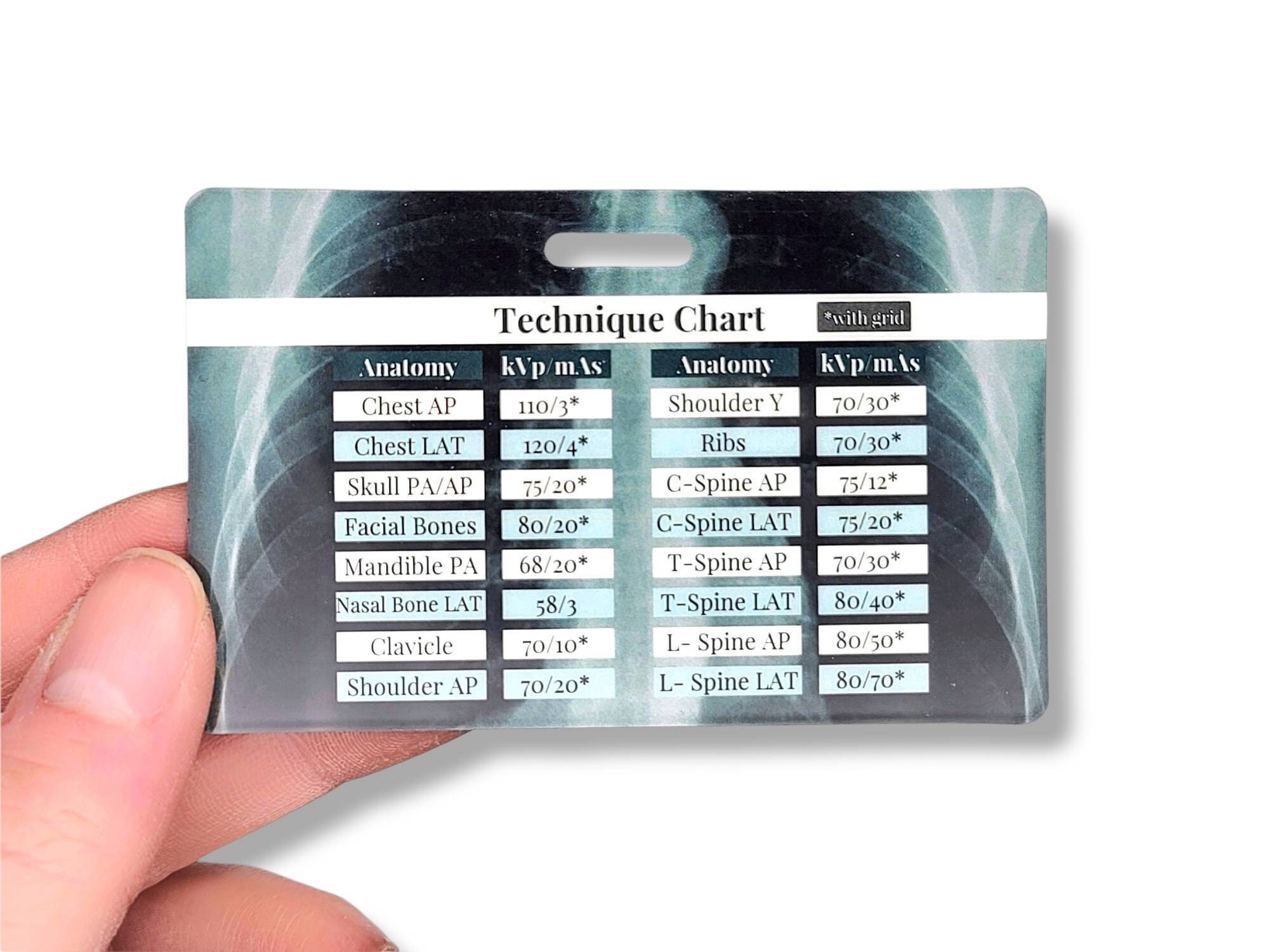
Human Kvp And Mas Technique Chart
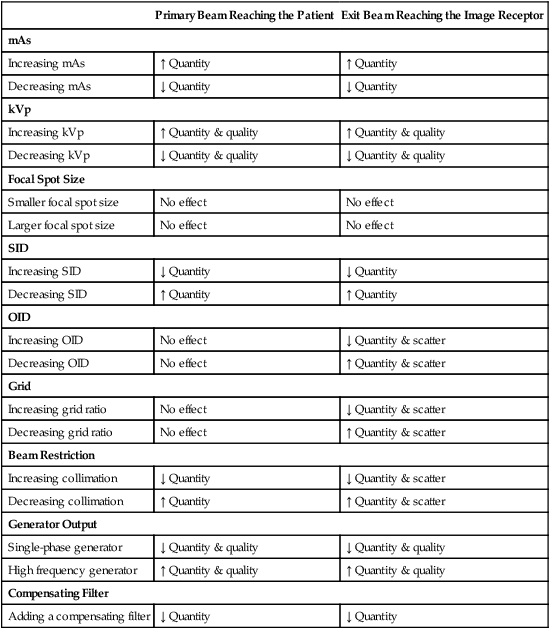
A Fixedkvp Technique Chart Uses Which of the Following GideonkruwBoyd

Xray Kvp And Mas Chart
Radiographic Technique Chart KVP Ma Time/S Sid Skull PDF
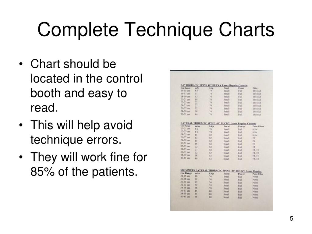
PPT Radiologic Technology Basic Cervical Radiography PowerPoint

Xray Technique Card Xray Mas and Kvp Chart Radiology Etsy

kVp/MAS Ranges for Radiology

Xray Technique Card Xray Mas and Kvp Chart Radiology Etsy Canada
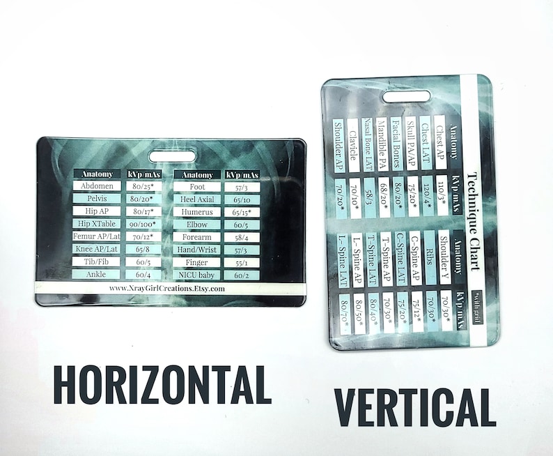
Xray Technique Card Xray Mas and Kvp Chart Radiology Etsy UK
Describe How The Photon Energy, Radiographic Contrast And Scale Of Contrast Vary As The Kvp Is Changed.
Keep In Mind When Making Your Choice For The Standard Kvp That Higher Kvp Will Allow You To Reduce The Mas And Reduce Patient Dose.
Web Make A Technique Chart.
Web 35 Cm X 43 Cm.
Related Post:
