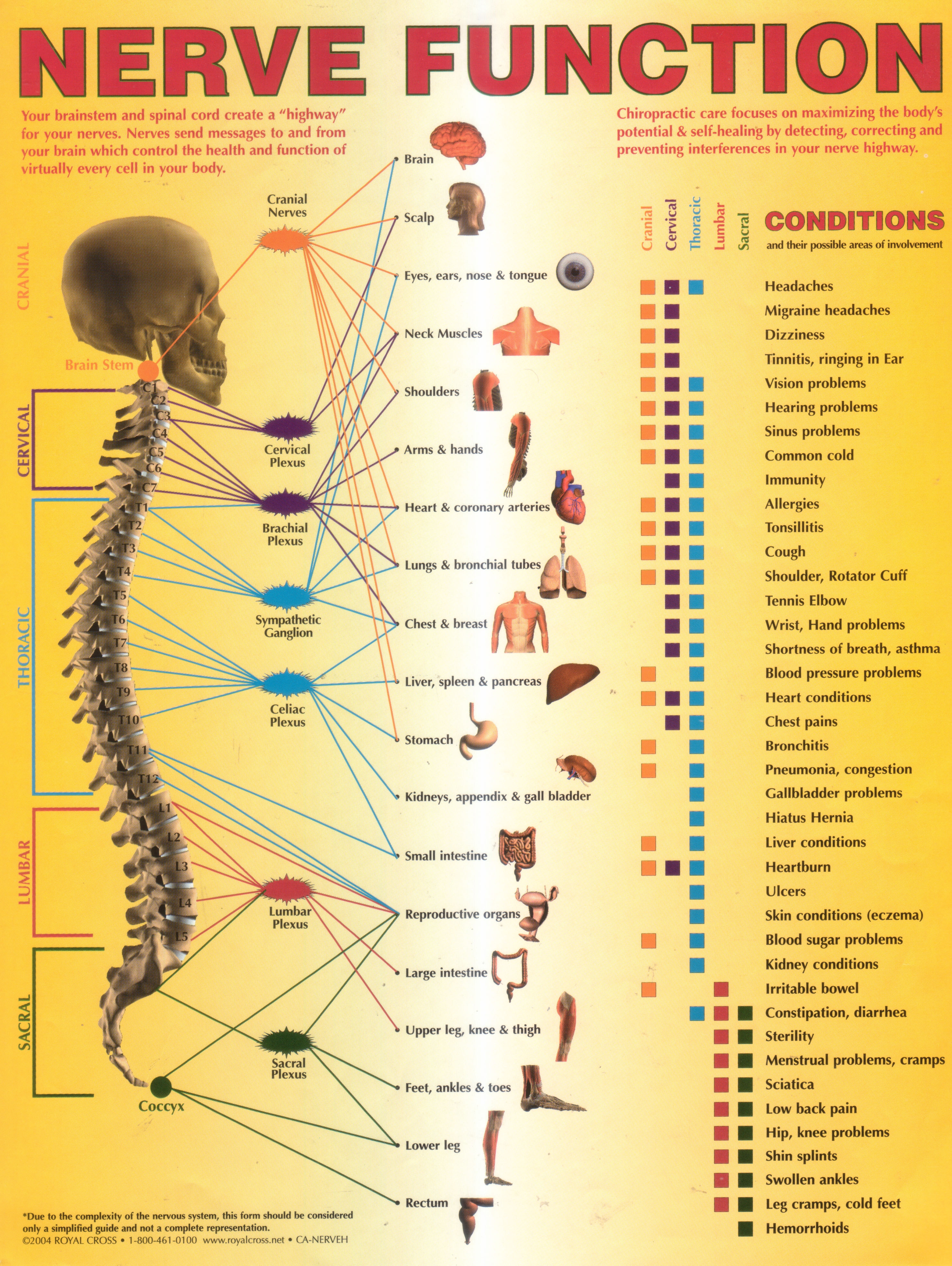
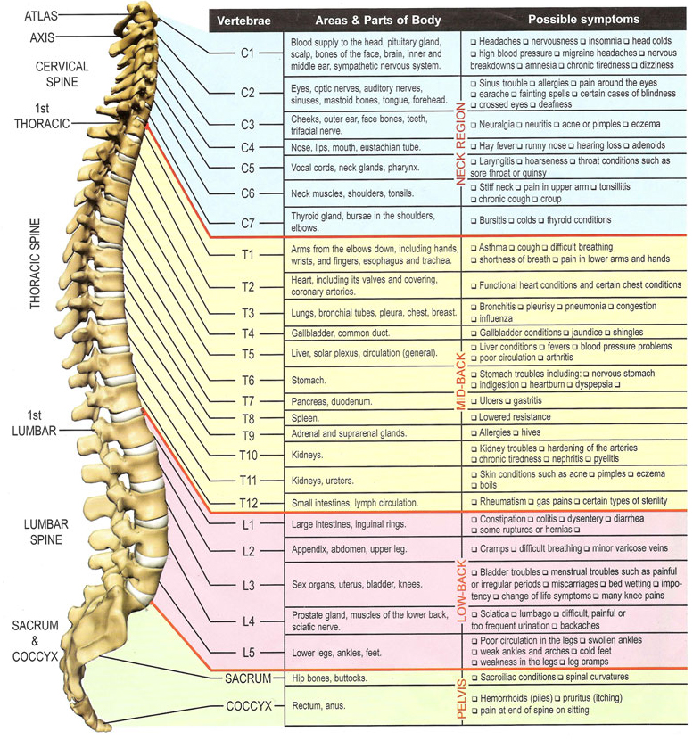
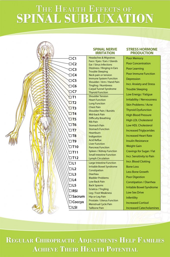
Nerve Innervation Chart
Nerve Innervation Chart - Web nerves of the upper limb. The sciatic nerve is a terminal branch of the sacral plexus. In general, the spinal cord consists of gray and white matter. All cranial nerves originate from nuclei in the brain. Clinically relevant nerves to the lower extremity. Also they transmit the motor commands from the cns to the muscles of the. A dermatome is an area of skin supplied by a single spinal nerve. Web we’ve created muscle anatomy charts for every muscle containing region of the body: Web the relevant anatomy of the innervation of the musculature of the back by the spinal nerves is centered around the lumbar spinal nerves, peripheral nerves of the lumbar plexus, spinal cord, and lumbar vertebral column. Web neurons, or nerve cell, are the main structural and functional units of the nervous system. Each chart groups the muscles of that region into its component groups, making your revision a million times easier. Web below is a chart that outlines the main functions of each of the spine nerve roots: The central system is the primary command center for the body, and is. Nerves of the lower limb. Its small branches innervate anterior thigh. View detailed diagrams of the brain, spinal cord, and other nervous system structures. Nerve cells are also called neurons. Web all innervate the lower limb and the pelvic region (figure 12.4.9 12.4. They are the structures through which the central nervous system (cns) receives sensory information from the periphery, and through which the activity of the trunk and the limbs. Web the relevant anatomy of the innervation of the musculature of the back by the spinal nerves is centered around the lumbar spinal nerves, peripheral nerves of the lumbar plexus, spinal cord, and lumbar vertebral column. Web afferent (sensory), efferent (motor), mixed. Web cranial nerves are the 12 nerves of the peripheral nervous system that emerge from the foramina and. It is important to mention that after the spinal nerves exit from the spine, they join together to form four paired clusters of. Web explore the nervous system with innerbody's interactive guide. It is formed from both anterior and posterior divisions of the anterior (ventral) rami of spinal nerves l4 through s3. In general, the spinal cord consists of gray. Web the somatic, voluntary, nervous system is responsible for providing sensory and motor innervation to skin, muscles and sensory organs. They include the olfactory nerve, which is. Each chart groups the muscles of that region into its component groups, making your revision a million times easier. Axillary, musculocutaneous, median, radial, and ulnar nerves. Nerves are like cables that carry electrical. Web below is a chart that outlines the main functions of each of the spine nerve roots: In this article, we break down the different types of nerves, as well. The cranial nerves are a set of twelve nerves that originate in the brain. Nerves of the upper limb. Clinically relevant nerves to the lower extremity. They also maintain certain autonomic functions like breathing, sweating or digesting food. Nerves of the upper limb. Web we’ve created muscle anatomy charts for every muscle containing region of the body: 9 and figure 12.4.10 12.4. There’s also a quick dermatomal map and myotomal chart for easy reference. Every neuron consists of a body (soma) and a number of processes (neurites). While the structure of a nerve is simple, their functions, innervations and nomenclature can be complex. Web nerves of the upper limb. There’s also a quick dermatomal map and myotomal chart for easy reference. Web the 30 dermatomes explained and located. Web its sensory fibers provide cutaneous innervation to the scalp, neck, chest, and axilla, as well as proprioceptive innervation of the same area via the lesser occipital nerve (c2 to c3), the great auricular nerve (c2, c3), transverse cervical nerve (c2, c3), and the supraclavicular nerve (c3, c4). Axillary, musculocutaneous, median, radial, and ulnar nerves. Web click on your choice. These impulses help you feel sensations and move your muscles. Web the relevant anatomy of the innervation of the musculature of the back by the spinal nerves is centered around the lumbar spinal nerves, peripheral nerves of the lumbar plexus, spinal cord, and lumbar vertebral column. There’s also a quick dermatomal map and myotomal chart for easy reference. The main. Web afferent (sensory), efferent (motor), mixed. Web the 30 dermatomes explained and located. While the structure of a nerve is simple, their functions, innervations and nomenclature can be complex. 9 and figure 12.4.10 12.4. Web click on your choice below: Web its sensory fibers provide cutaneous innervation to the scalp, neck, chest, and axilla, as well as proprioceptive innervation of the same area via the lesser occipital nerve (c2 to c3), the great auricular nerve (c2, c3), transverse cervical nerve (c2, c3), and the supraclavicular nerve (c3, c4). Web the somatic, voluntary, nervous system is responsible for providing sensory and motor innervation to skin, muscles and sensory organs. Web knowing muscle innervations is an important skill for medical professionals such as physical therapists, physicians, and occupational therapists. Web explore the nervous system with innerbody's interactive guide. The nerve cell body contains the cellular organelles and is where neural impulses (action potentials) are generated. Also they transmit the motor commands from the cns to the muscles of the. It is formed from both anterior and posterior divisions of the anterior (ventral) rami of spinal nerves l4 through s3. The central system is the primary command center for the body, and is. Web muscle innervation reference tables. The sciatic nerve is a terminal branch of the sacral plexus. There’s also a quick dermatomal map and myotomal chart for easy reference.
Annual World Spine Day Campaign Nerve Function Chart

Nervous System Anatomy Posters Set of 6 Etsy Nervous system anatomy

Nerve compression causes, symptoms, diagnosis & treatment

Anatomy Anatomy Of The Spinal Cord And Nerves Youtube vrogue.co

Upper Extremity Nerve Innervation Chart

Spinal Nerve Function Anatomical Chart Anatomy Models and Anatomical

Lumbar Spinal Nerve Chart

upper extremity peripheral nerves netter Google Search Thần kinh

Upper Limbs Nervous System Poster Anterior Chartex

Nerve Chart Hunter Chiropractic Wellness Centre
Web We’ve Created Muscle Anatomy Charts For Every Muscle Containing Region Of The Body:
Web The Relevant Anatomy Of The Innervation Of The Musculature Of The Back By The Spinal Nerves Is Centered Around The Lumbar Spinal Nerves, Peripheral Nerves Of The Lumbar Plexus, Spinal Cord, And Lumbar Vertebral Column.
Nerve Cells Are Also Called Neurons.
Its Small Branches Innervate Anterior Thigh Muscles And Receives Sensory Information From The Anterior And Medial Aspects Of The Thigh.
Related Post: