Nerve Spine Chart
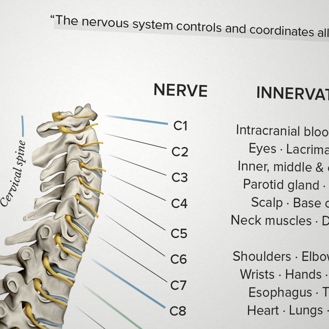
Nerve Spine Chart - Web the spine’s four sections, from top to bottom, are the cervical (neck), thoracic (abdomen,) lumbar (lower back), and sacral (toward tailbone). After exiting the vertebral column, the spinal nerves travel to their target areas, which can include muscles, skin, and other tissues. Your spinal cord is a column of nerves that travels through your spinal canal. Web the spine is divided into four regions which contain vertebrae: The cord extends from your skull to your lower back. Each nerve forms from nerve fibers, known as fila radicularia, extending from the posterior (dorsal) and anterior (ventral) roots of the spinal cord. Web there are 31 pairs of spinal nerves: Numbers indicate the types of nerve fibers: It provides a visual representation of the anatomy and physiology of the nervous system, which helps to explain the function of the spinal nerves. The roots connect via interneurons. Web spinal nerves are all mixed nerves with both sensory and motor fibers. Eight cervical spinal nerves on each side of the spine called c1 through c8. L2, l3, and l4 spinal nerves provide sensation to the front part of the thigh and inner side of the lower leg. After exiting the vertebral column, the spinal nerves travel to their. The dorsal root is the afferent sensory root and carries sensory information to the brain. The back functions are many, such as to house and protect the spinal cord, hold the body and head upright, and adjust the movements of the upper. Web l1 spinal nerve provides sensation to the groin and genital regions and may contribute to the movement. Web the spine is divided into four regions which contain vertebrae: Twelve thoracic spinal nerves in each side of the body called t1 through t12. The back is the body region between the neck and the gluteal regions. Web the spine’s four sections, from top to bottom, are the cervical (neck), thoracic (abdomen,) lumbar (lower back), and sacral (toward tailbone).. Web many nerves come from the spinal cord, pass through foramina (holes) formed by notches of 24 vertebrae in the movable spinal column, and innervate or supply specific areas and parts of the body.2 whenever specific areas or parts of the body are malfunctioning, generalized and/or specific symptoms are possible.3. L2, l3 and l4 spinal nerves provide sensation to the. You have 31 spinal nerves and 30 dermatomes. L2, l3, and l4 spinal nerves provide sensation to the front part of the thigh and inner side of the lower leg. Web the nerve roots are numbered based on their location in the spinal cord, with the first cervical nerve root (c1) located at the top of the spinal cord near. Web the spine’s four sections, from top to bottom, are the cervical (neck), thoracic (abdomen,) lumbar (lower back), and sacral (toward tailbone). The exact area that each dermatome covers can be different from person. It is important to mention that after the spinal nerves exit from the spine, they join together to form four paired clusters of. Five lumbar spinal. Web 1 coccygeal (co1) route: Each spinal nerve is a mixed nerve, formed from the combination of nerve root fibers from its dorsal and ventral roots. Hover over each part to see what they do. Web there are 31 pairs of spinal nerves: The exact area that each dermatome covers can be different from person. Numbers indicate the types of nerve fibers: Thoracic spinal nerves are not part of any plexus, but give rise to the intercostal nerves directly. Web l1 spinal nerve provides sensation to your groin and genital area and helps move your hip muscles. Eight cervical spinal nerve pairs, 12 thoracic pairs , five lumbar pairs, five sacral pairs, and one coccygeal.. Web below is a chart that outlines the main functions of each of the spine nerve roots: Your spinal cord is a column of nerves that travels through your spinal canal. These nerves carry messages between your brain and muscles. Web there are 31 bilateral pairs of spinal nerves, named from the vertebra they correspond to. Web spine nerves anatomy,. For the most part, the spinal nerves exit the vertebral canal through the intervertebral foramen below their corresponding vertebra. The cord extends from your skull to your lower back. 8 cervical, 12 thoracic, 5 lumbar, 5 sacral, and 1 coccygeal, named according to their corresponding vertebral levels. L2, l3, and l4 spinal nerves provide sensation to the front part of. Each spinal nerve is a mixed nerve, formed from the combination of nerve root fibers from its dorsal and ventral roots. Eight cervical spinal nerves on each side of the spine called c1 through c8. These nerves carry messages between your brain and muscles. Web spinal nerves are all mixed nerves with both sensory and motor fibers. The dorsal root is the afferent sensory root and carries sensory information to the brain. Web spinal nerves are mixed nerves that interact directly with the spinal cord to modulate motor and sensory information from the body’s periphery. For the most part, the spinal nerves exit the vertebral canal through the intervertebral foramen below their corresponding vertebra. It is important to mention that after the spinal nerves exit from the spine, they join together to form four paired clusters of. Web dermatomes are areas of skin that are connected to a single spinal nerve. Web spine nerves anatomy, diagram & function | body maps. The exact area that each dermatome covers can be different from person. The peripheral nerves are responsible for sensations and muscle movements. The vertebral column’s most important physiologic function is protecting the spinal cord,. Web 5 sacral spinal nerves. The cervical, the thoracic, the lumbar, and the sacral. Numbers indicate the types of nerve fibers:
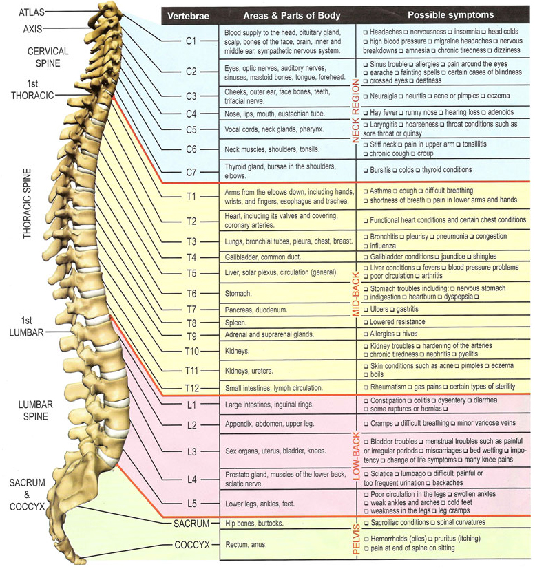
Vertebral Subluxation & Nerve Chart Healing Hands Chiropractic

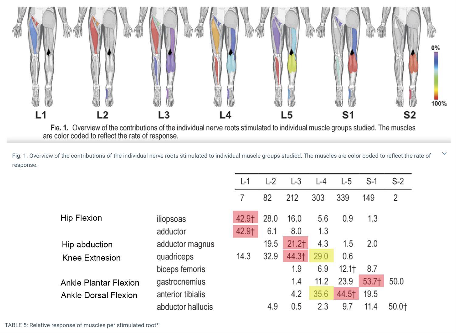
Lumbar Spinal Nerve Chart

Spinal Nerve Chart Pain

Pain Spine Nerve Chart

Spinal Nerve Chart medschool doctor medicalstudent Image Credits

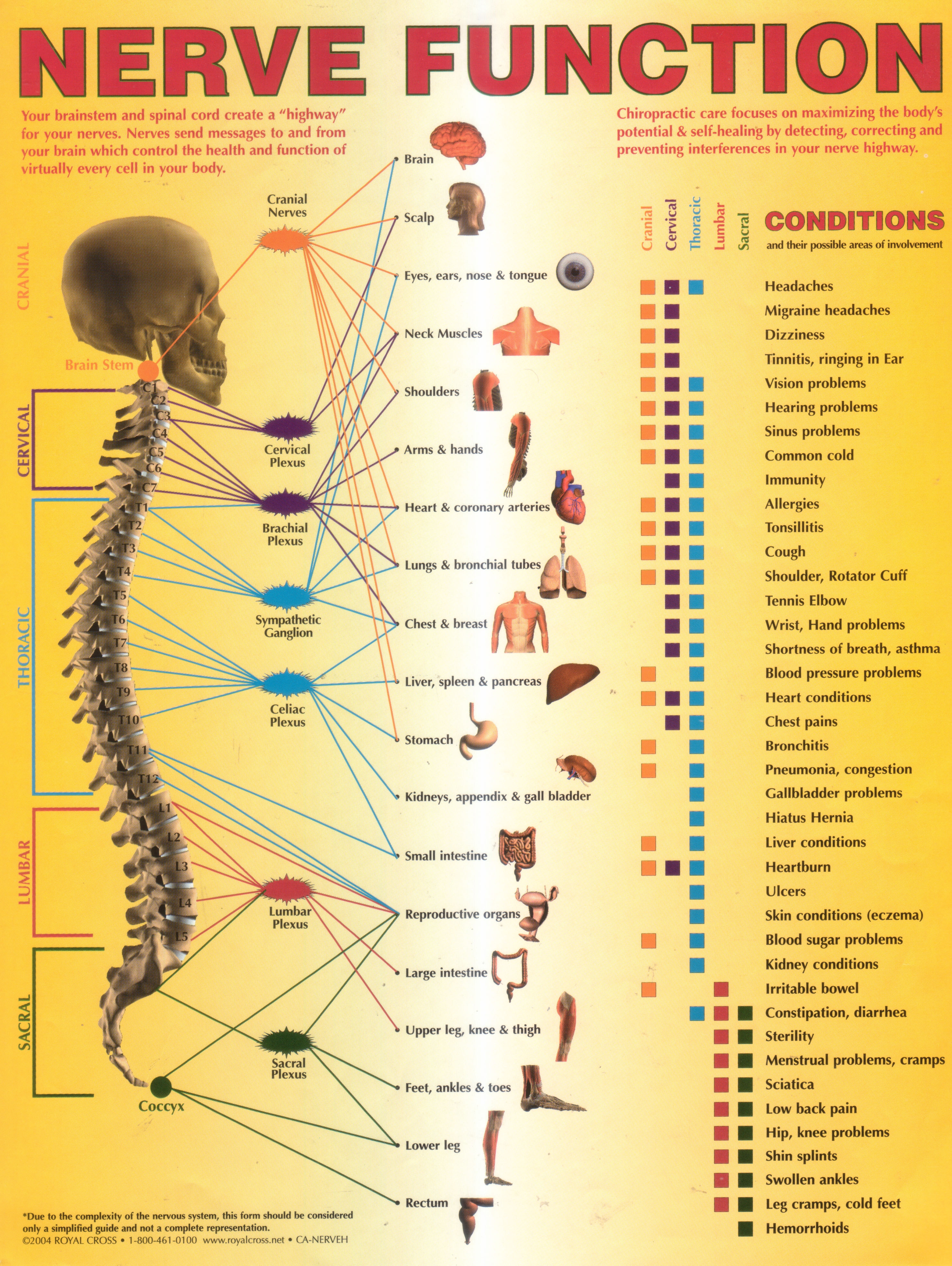
Annual World Spine Day Campaign Nerve Function Chart

the spiral nerve function is shown in this manual for students to learn

Spinal Nerve Function Anatomical Chart Anatomy Models and Anatomical

Lumbar Spinal Nerve Chart

pelota equivocado telegrama spinal cord nerves anatomy De Verdad Ya que
For Most Spinal Segments, The Nerve Roots Run Through The Bony Canal, And At Each Level A Pair Of Nerve Roots Exits From The Spine.
Hover Over Each Part To See What They Do.
You Have 31 Spinal Nerves And 30 Dermatomes.
Web The Nerve Roots Are Numbered Based On Their Location In The Spinal Cord, With The First Cervical Nerve Root (C1) Located At The Top Of The Spinal Cord Near The Base Of The Brain, And The Last Sacral Nerve Root (S5) Located At The Bottom.
Related Post: