Nuchal Fold Thickness Measurement Chart
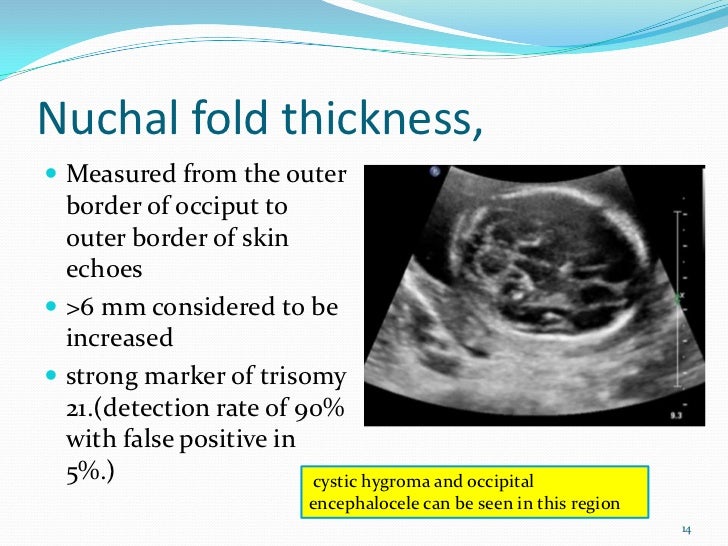
Nuchal Fold Thickness Measurement Chart - Web the nuchal fold is a normal fold of skin seen at the back of the fetal neck during the second trimester of pregnancy. Web nuchal fold thickness is measured towards the end of the second trimester. [1,2] an nf measurement greater than 5 mm at 14 to 17 +6 weeks of gestation [3,4] or 6 mm at 18 to 28 weeks of gestation has been. Thickness is independent of maternal age. It is part of the combined screening test for down's syndrome, which is offered at around 12 weeks of pregnancy. It is examined using ultrasound as part of combined screening for down, edwards and patau syndromes from 11 to 13 weeks and six days of pregnancy. In a small number of pregnancies a thick nuchal fold is found. Web nuchal fold can be spuriously thickened by angling caudally (intersecting the inferior level of the cerebellum and occiput). Pt ratio could help in differentiating euploid and down syndrome fetuses. Web a nuchal translucency (nt) scan is an ultrasound scan to measure the amount of fluid at the back of your baby's neck. It can be a normal variation or associated with extra fluid in the skin (oedema) which may be due to an infection or a chromosomal condition such as downs’s syndrome (trisomy 21). Web this leaflet is to help you understand what nuchal translucency (nt) is, what tests you need, and the implication of being diagnosed for you, your baby, and. Web increased measurement of the nuchal fold (≥ 6 mm from 14 weeks to 22 weeks of gestational age) is considered a soft marker for chromosomal aneuplodies, as well as for structural defects in the fetus, most commonly cardiac defects. Web most authors use a thickness of ³ 3 mm to define abnormal (some authors use 2.5mm). Thickness is independent. Web this leaflet is to help you understand what nuchal translucency (nt) is, what tests you need, and the implication of being diagnosed for you, your baby, and your family. Web the measurement of nuchal fold (nf) thickness during the second trimester is considered to be one of the most sensitive and specific isolated ultrasound marker for the identification of. Thickness is independent of maternal age. We also describe the outcome of fetuses with increased second trimester nuchal folds. It is part of the combined screening test for down's syndrome, which is offered at around 12 weeks of pregnancy. [1,2] an nf measurement greater than 5 mm at 14 to 17 +6 weeks of gestation [3,4] or 6 mm at. It is examined using ultrasound as part of combined screening for down, edwards and patau syndromes from 11 to 13 weeks and six days of pregnancy. Sonographic assessment of fetal nuchal skin fold thickness (nft) was first described by benacerraf and colleagues in 19851. Web the nuchal fold normally measures less than 6mm at 20 weeks. It can be a. Web nuchal fold thickness is measured towards the end of the second trimester. Web abstract objective to assess the effect of imaging angle and fetal presentation on the measurement of nuchal skin fold thickness (nft) in the second trimester. We also describe the outcome of fetuses with increased second trimester nuchal folds. Web increased measurement of the nuchal fold (≥. Web the nuchal fold normally measures less than 6mm at 20 weeks. This is an area of tissue at the back of an unborn baby's neck. Web nuchal fold thickness is measured towards the end of the second trimester. It is examined using ultrasound as part of combined screening for down, edwards and patau syndromes from 11 to 13 weeks. Web the nuchal fold is thick when it measures 6 mm or more. Web the measurement of nuchal fold (nf) thickness during the second trimester is considered to be one of the most sensitive and specific isolated ultrasound marker for the identification of suspected cases of trisomy 21. [ 1, 2] an nf measurement greater than 5 mm at 14. Web the nuchal translucency test measures the nuchal fold thickness. The measurement is indicated by the calipers. Sonographic assessment of fetal nuchal skin fold thickness (nft) was first described by benacerraf and colleagues in 19851. Web measuring nuchal skin fold thickness after tilting the transducer towards the occiput approximately 30° while maintaining the appropriate intracranial landmarks. The results of your. The formula is as follows: As nuchal translucency size increases, the chances of a chromosomal abnormality and mortality increase; Web the nuchal translucency test measures the nuchal fold thickness. Web the nuchal fold normally measures less than 6mm at 20 weeks. Web the measurement of nuchal fold (nf) thickness during the second trimester is considered to be one of the. Increased thickness of the nuchal fold is a soft marker associated with multiple fetal anomalies, and is measured on a routine second trimester ultrasound. Pt ratio could help in differentiating euploid and down syndrome fetuses. Methods fetal nft was prospectively m. Thickness is independent of maternal age. Web the measurement of nuchal fold (nf) thickness during the second trimester is considered to be one of the most sensitive and specific isolated ultrasound marker for the identification of suspected cases of trisomy 21. Measuring this thickness helps assess the risk for down syndrome and other genetic problems in the baby. Web the objective of the study is to investigate the effect of nuchal fold (nf) in predicting fetal growth restriction (fgr) using machine learning (ml), to explain the model's results using. Web the nuchal fold is a normal fold of skin seen at the back of the fetal neck during the second trimester of pregnancy. We also describe the outcome of fetuses with increased second trimester nuchal folds. In a small number of pregnancies a thick nuchal fold is found. Web the nuchal fold (nf) thickness is a measurement performed on prenatal ultrasound, and is the distance from the outer edge of the occipital bone to the outer edge of the skin in the midline. Web the nuchal translucency test measures the nuchal fold thickness. The results of your nt scan are combined with blood test results and other factors, such as your age. 65% of the largest translucencies (>6.5mm) are due to chromosomal abnormality, while fatality is 19% at this size. Web the nuchal fold is thick when it measures 6 mm or more. Web this leaflet is to help you understand what nuchal translucency (nt) is, what tests you need, and the implication of being diagnosed for you, your baby, and your family.
Comparison of nuchal fold thickness measurements by twoand

Nuchal Fold Measurement Chart A Visual Reference of Charts Chart Master

Measurement of nuchal skin fold thickness in the second trimester

Comparison of nuchal fold thickness measurements by twoand

Nuchal Fold Thickness Normal or abnormal? Expert insights in 2023

Level II usg

Increased Fetal Nuchal Translucency Thickness and Normal Karyotype

Nuchal fold (NF) values in euploid fetuses. Download Scientific Diagram

What is the Nuchal Fold Measurement?

Table 2 from Reference values of nuchal translucency thickness in a
Does A Thick Nuchal Fold Mean The Baby Has A Problem?
The Measurement Is Indicated By The Calipers.
The Formula Is As Follows:
This Is An Area Of Tissue At The Back Of An Unborn Baby's Neck.
Related Post: