Spine Chart With Nerves
Spine Chart With Nerves - Web below is a chart that outlines the main functions of each of the spine nerve roots: Spinal nerves are mixed nerves that emerge from the spinal cord and carry both motor and sensory information between the spinal cord and various parts of the body. The dorsal root is the afferent sensory root and carries sensory information to the brain. We provide a pictorial review of neuroradiological. Your lumbar spine connects to your pelvis and bears most of your body’s weight, as well as the stress of lifting and carrying items. Many of the nerves of the peripheral nervous system, or pns, branch out from the spinal cord. The cervical, the thoracic, the lumbar, and the sacral. Each spinal nerve is a mixed nerve, formed from the combination of nerve root fibers from its dorsal and ventral roots. Web the arthritis foundation states that 31 pairs of nerves branch off the spinal cord to other parts of the body. Type 1 neurofibromatosis (nf1) is the most common neurocutaneous disorder, and it is an inherited condition that causes a tumour predisposition. These complex networks of nerves enable the brain to receive sensory inputs from the skin and to send motor controls for muscle movements. Your spinal cord is a column of nerves that travels through your spinal canal. The back is the body region between the neck and the gluteal regions. Central nervous system (cns) manifestations are a significant cause of. Web spinal nerves are mixed nerves that interact directly with the spinal cord to modulate motor and sensory information from the body’s periphery. Thoracic spinal nerves are not part of any plexus, but give rise to the intercostal nerves directly. Your lumbar spine connects to your pelvis and bears most of your body’s weight, as well as the stress of. We provide a pictorial review of neuroradiological. Web spinal nerves are mixed nerves that interact directly with the spinal cord to modulate motor and sensory information from the body’s periphery. The spinal cord transmits signals from the brain to the rest of a person’s body. The cervical, the thoracic, the lumbar, and the sacral. The roots connect via interneurons. Your spinal nerves help to relay sensory, motor, and autonomic information between the rest of. Web spinal nerves are all mixed nerves with both sensory and motor fibers. Web learn the anatomy of the spinal nerves, including their roots, components and functions faster and more efficiently with this comprehensive article. The cervical portion of the spine is an important one. It is part of the axial skeleton and extends from the base of the skull to the tip of the coccyx. The roots connect via interneurons. It comprises the vertebral column (spine) and two compartments of back muscles; Web spinal nerves are all mixed nerves with both sensory and motor fibers. The peripheral nerves are responsible for sensations and muscle. The dorsal root is the afferent sensory root and carries sensory information to the brain. On the chart below you will see 4 columns (vertebral level, nerve root, innervation, and possible symptoms). Web the spine is divided into four regions which contain vertebrae: Web below is a chart that outlines the main functions of each of the spine nerve roots:. Web spinal nerves are all mixed nerves with both sensory and motor fibers. Web how to use the spinal nerve chart: Web explore the anatomy and functions of lumbar spinal nerves. It is important to mention that after the spinal nerves exit from the spine, they join together to form four paired clusters of. Web there are 31 pairs of. The spinal cord begins at the base of the brain and extends into the pelvis. Central nervous system (cns) manifestations are a significant cause of morbidity and mortality in nf1. Your spinal nerves help to relay sensory, motor, and autonomic information between the rest of. The back is the body region between the neck and the gluteal regions. Web the. Together, the brain and spinal cord make up the central nervous system. Web the spinal cord and its nerves are the means by which the body and brain communicate with one another. The cervical portion of the spine is an important one anatomically and clinically. Web there are 31 pairs of nerves that emerge from the spine. On the chart. The roots connect via interneurons. The spinal cord begins at the base of the brain and extends into the pelvis. These nerves carry messages between your brain and muscles. Web below is a chart that outlines the main functions of each of the spine nerve roots: The cervical portion of the spine is an important one anatomically and clinically. Web spinal nerves are all mixed nerves with both sensory and motor fibers. Web spinal cord and nerves: The back is the body region between the neck and the gluteal regions. A long, tubular bundle of nerves that extends from the brainstem down the vertebral column, protected by fluid in the spinal canal and surrounded by ligaments and bone for protection. Web how to use the spinal nerve chart: Your lumbar spine connects to your pelvis and bears most of your body’s weight, as well as the stress of lifting and carrying items. Web there are 31 pairs of nerves that emerge from the spine. The cord extends from your skull to your lower back. Eight cervical spinal nerve pairs, 12 thoracic pairs , five lumbar pairs, five sacral pairs, and one coccygeal. If injured, therapy or surgery can help. Spinal nerves are mixed nerves that emerge from the spinal cord and carry both motor and sensory information between the spinal cord and various parts of the body. Web learn the anatomy of the spinal nerves, including their roots, components and functions faster and more efficiently with this comprehensive article. Central nervous system (cns) manifestations are a significant cause of morbidity and mortality in nf1. Web the vertebral column (spine or backbone) is a curved structure composed of bony vertebrae that are interconnected by cartilaginous intervertebral discs. The spinal cord transmits signals from the brain to the rest of a person’s body. We provide a pictorial review of neuroradiological.
Spinal Nerves Anatomical Chart Spine and Cranial Nervous System
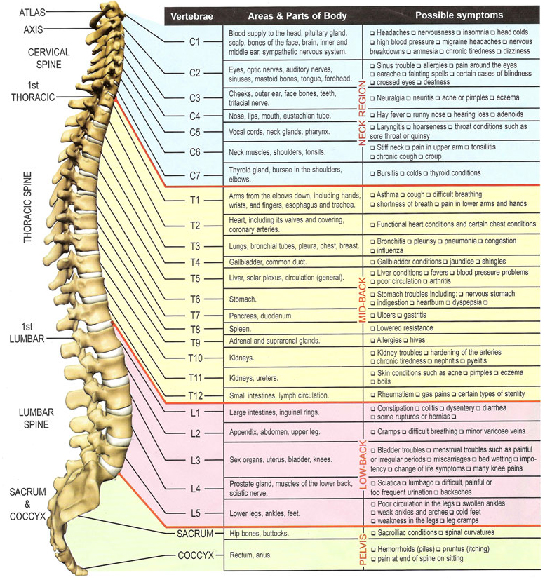
spine and nerve chart
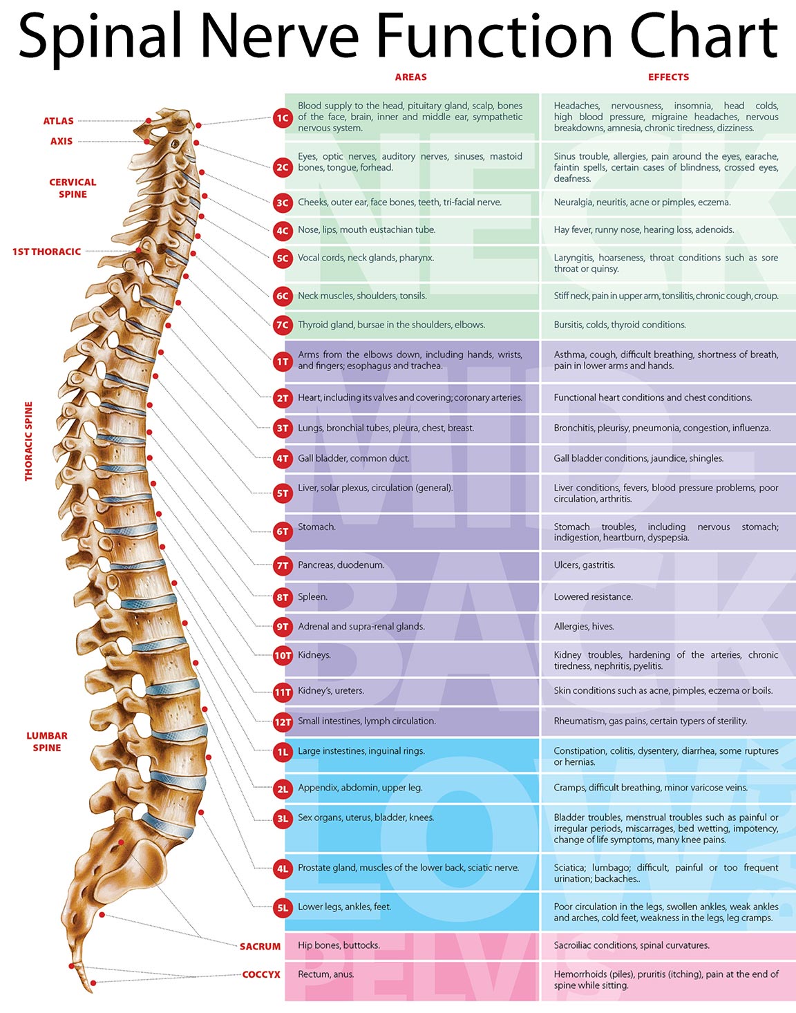
The Spinal Nerves Chart

Spine Chart ANDERSON CHIROPRACTIC

Spinal Nerve Chart medschool doctor medicalstudent Image Credits

Lumbar Spinal Nerve Chart
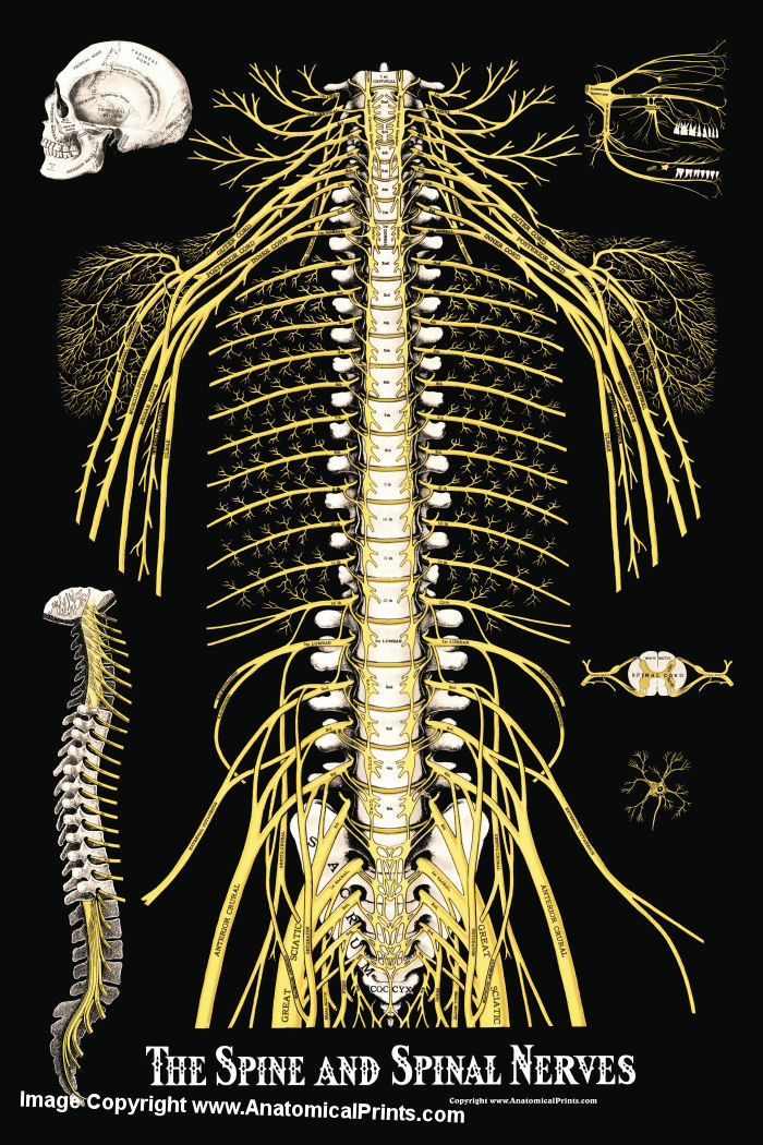
The Spine and Spinal Nerves Poster Clinical Charts and Supplies
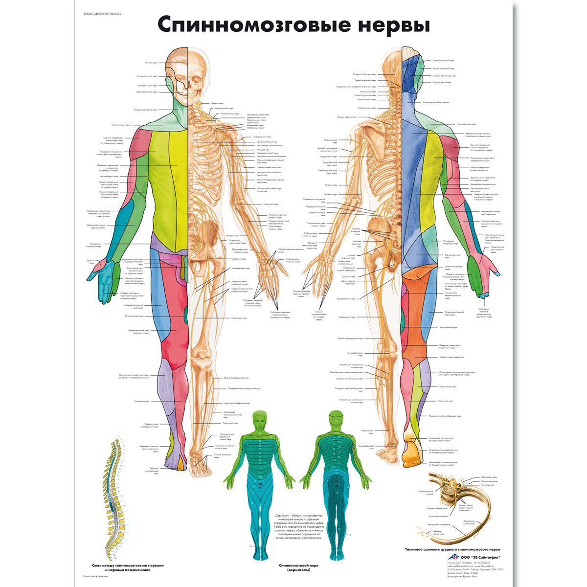
Spinal Nerves Chart 1002329 VR6621L 3B Scientific ZVR6621L

Spinal Nerve Function Anatomical Chart Anatomy Models and Anatomical
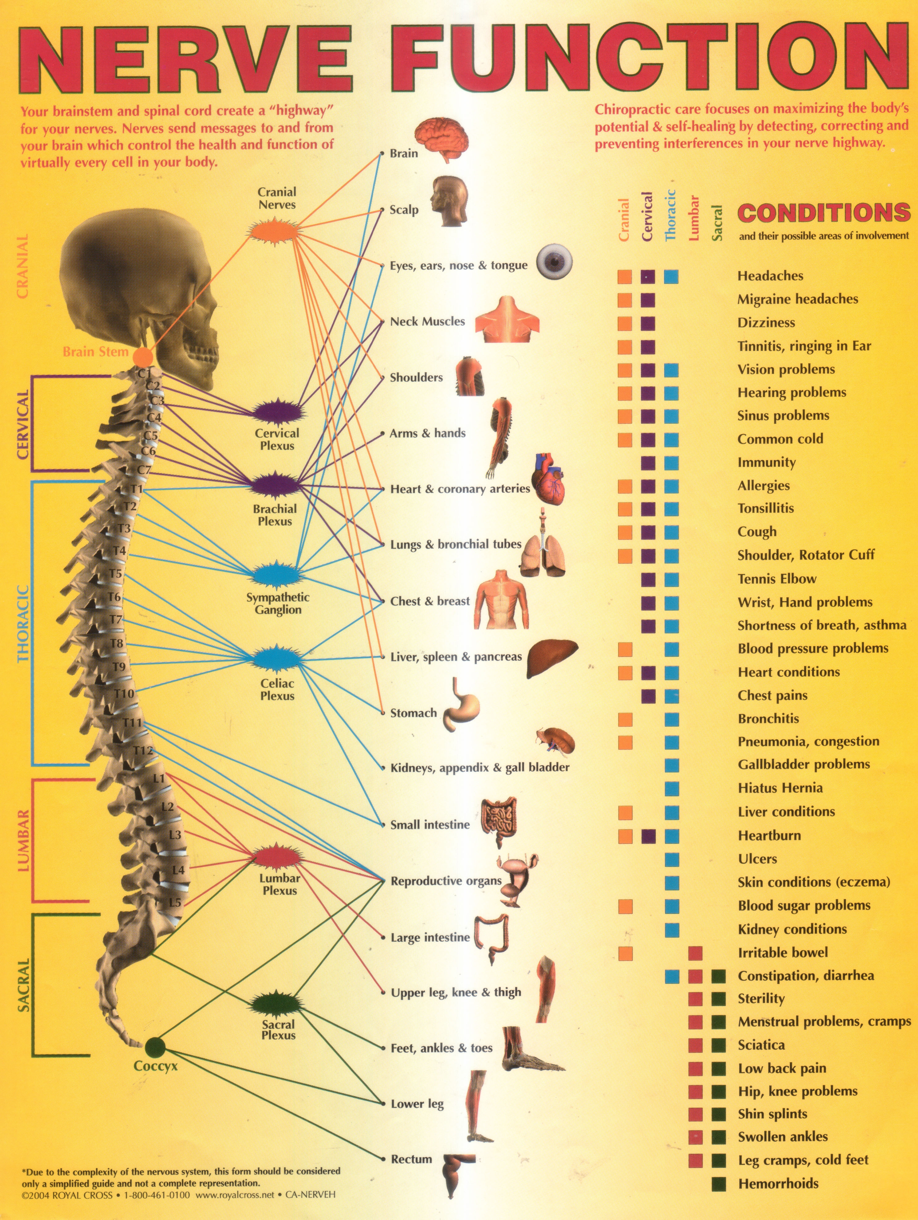
Annual World Spine Day Campaign Nerve Function Chart
The Cervical, The Thoracic, The Lumbar, And The Sacral.
The Dorsal Root Is The Afferent Sensory Root And Carries Sensory Information To The Brain.
Web Your Lumbar Spine Supports The Upper Two Sections Of Your Spine — The Seven Vertebrae In Your Neck (Cervical Spine) And 12 Vertebrae In Your Chest (Thoracic Spine) — And The Weight Of Your Head.
Web Explore The Anatomy And Functions Of Lumbar Spinal Nerves.
Related Post: