The Spinal Nerves Chart
The Spinal Nerves Chart - The central illustration shows a posterior view of the spinal nerves exiting from the vertebral column and running throughout the body. The spinal cord serves as the central pathway for transmitting sensory and motor signals between the brain and the body through specific. Thomas scioscia, md , orthopedic surgeon. The spinal nerves innervate the skin and the muscles of a specific region. Central nervous system (cns) manifestations are a significant cause of morbidity and mortality in nf1. The spinal nerves send sensory messages to the sensory roots, then to sensory fibers in the posterior (back or dorsal) part of the spinal cord. Ventral root (located in front) that carries motor signals from the brain to that nerve root’s myotome, which is the group of muscles that it controls. Web the spinal cord and peripheral nerves. Web your spinal column or ‘backbone’ is made up of 24 vertebrae: Web spinal nerves are peripheral nerves that emerge from the spinal cord and carry motor, sensory, and autonomic signals between the spinal cord and the rest of the body. This spinal nerves anatomical chart illustrates spinal nerves, cranial nerves and diagrams the portion of the thoracic spinal cord with spinal nerves. Eight cervical spinal nerve pairs, 12 thoracic pairs , five lumbar pairs, five sacral pairs, and one coccygeal. The spinal nerves send sensory messages to the sensory roots, then to sensory fibers in the posterior (back or dorsal). Lumbar spinal nerves carry sensory and motor information to the lower body. It is important to mention that after the spinal nerves exit from the spine, they join together to form four paired clusters of. Thoracic spinal nerves are not part of any plexus, but give rise to the intercostal nerves directly. On the chart below you will see 4. 8 cervical, 12 thoracic, 5 lumbar, 5 sacral, and 1 coccygeal, named according to their corresponding vertebral levels. The spinal nerves anatomical chart also shows spinal cord segments, cutaneous distribution of spinal nerves and dermal segmentation. Type 1 neurofibromatosis (nf1) is the most common neurocutaneous disorder, and it is an inherited condition that causes a tumour predisposition. 8 cervical, 12. 8 cervical, 12 thoracic, 5 lumbar, 5 sacral, and 1 coccygeal, named according to their corresponding vertebral levels. Your spinal cord, made up of billions of nerves, lies inside. A dermatome is a specific area of skin that is supplied by. Each spinal nerve is a mixed nerve, formed from the combination of nerve root fibers from its dorsal and. Web below is a chart that outlines the main functions of each of the spine nerve roots: On the chart below you will see 4 columns (vertebral level, nerve root, innervation, and possible symptoms). Web a very popular and useful chart, the spinal nerves illustrates the spinal nerves and pathways through the body. Spinal nerves are mixed nerves that emerge. Web learn the anatomy of the spinal nerves, including their roots, components and functions faster and more efficiently with this comprehensive article. Web below is a chart that outlines the main functions of each of the spine nerve roots: Lumbar spinal nerves carry sensory and motor information to the lower body. 8 cervical, 12 thoracic, 5 lumbar, 5 sacral, and. Web each pair of spinal nerves roughly correspond to a segment of the vertebral column: Web health library / body systems & organs / spine structure and function. Your spine is made up of vertebrae (bones), disks, joints, soft tissues, nerves and your spinal cord. Web effect of tregs on the recovery of neurological symptoms in eae mice. Web your. Central nervous system (cns) manifestations are a significant cause of morbidity and mortality in nf1. Your spine is an important bone structure that supports your body and helps you walk, twist and move. Web spinal nerves are peripheral nerves that emerge from the spinal cord and carry motor, sensory, and autonomic signals between the spinal cord and the rest of. Spinal nerves emerge from the spinal cord and reorganize through plexuses, which then give rise to systemic nerves. Web medically reviewed by anatomy team. Your spinal cord, made up of billions of nerves, lies inside. It is important to mention that after the spinal nerves exit from the spine, they join together to form four paired clusters of. Each spinal. Web how to use the spinal nerve chart: The point at which a nerve exits the spinal cord is called a nerve root. Eight cervical spinal nerve pairs, 12 thoracic pairs , five lumbar pairs, five sacral pairs, and one coccygeal. Web health library / body systems & organs / spine structure and function. On the chart below you will. The dorsal root is the afferent sensory root and carries sensory information to the brain. It is important to mention that after the spinal nerves exit from the spine, they join together to form four paired clusters of. Web spinal nerves are peripheral nerves that emerge from the spinal cord and carry motor, sensory, and autonomic signals between the spinal cord and the rest of the body. 8 cervical, 12 thoracic, 5 lumbar, 5 sacral, and 1 coccygeal. Web below is a chart that outlines the main functions of each of the spine nerve roots: Important skeletal structures are included. There are 31 pairs of spinal nerves: The peripheral nerves are responsible for sensations and muscle movements. We provide a pictorial review of neuroradiological. Central nervous system (cns) manifestations are a significant cause of morbidity and mortality in nf1. Spinal cord and spinal nerve roots. The spinal cord starts at the base of the brain, runs throughout the cervical and thoracic spine, and typically ends at the lower part of the thoracic spine. These nerves are essential for transmitting sensory signals to the brain and for carrying motor commands from the brain to muscles. On the chart below you will see 4 columns (vertebral level, nerve root, innervation, and possible symptoms). Eight cervical spinal nerve pairs, 12 thoracic pairs , five lumbar pairs, five sacral pairs, and one coccygeal. Your spine is an important bone structure that supports your body and helps you walk, twist and move.
Spinal Nerves What They Are and What They Do

Spinal Nerve Function Anatomical Chart Anatomy Models and Anatomical

Spinal Nerve Function Cheat Sheet Spinal nerve, Nerves function and Chart
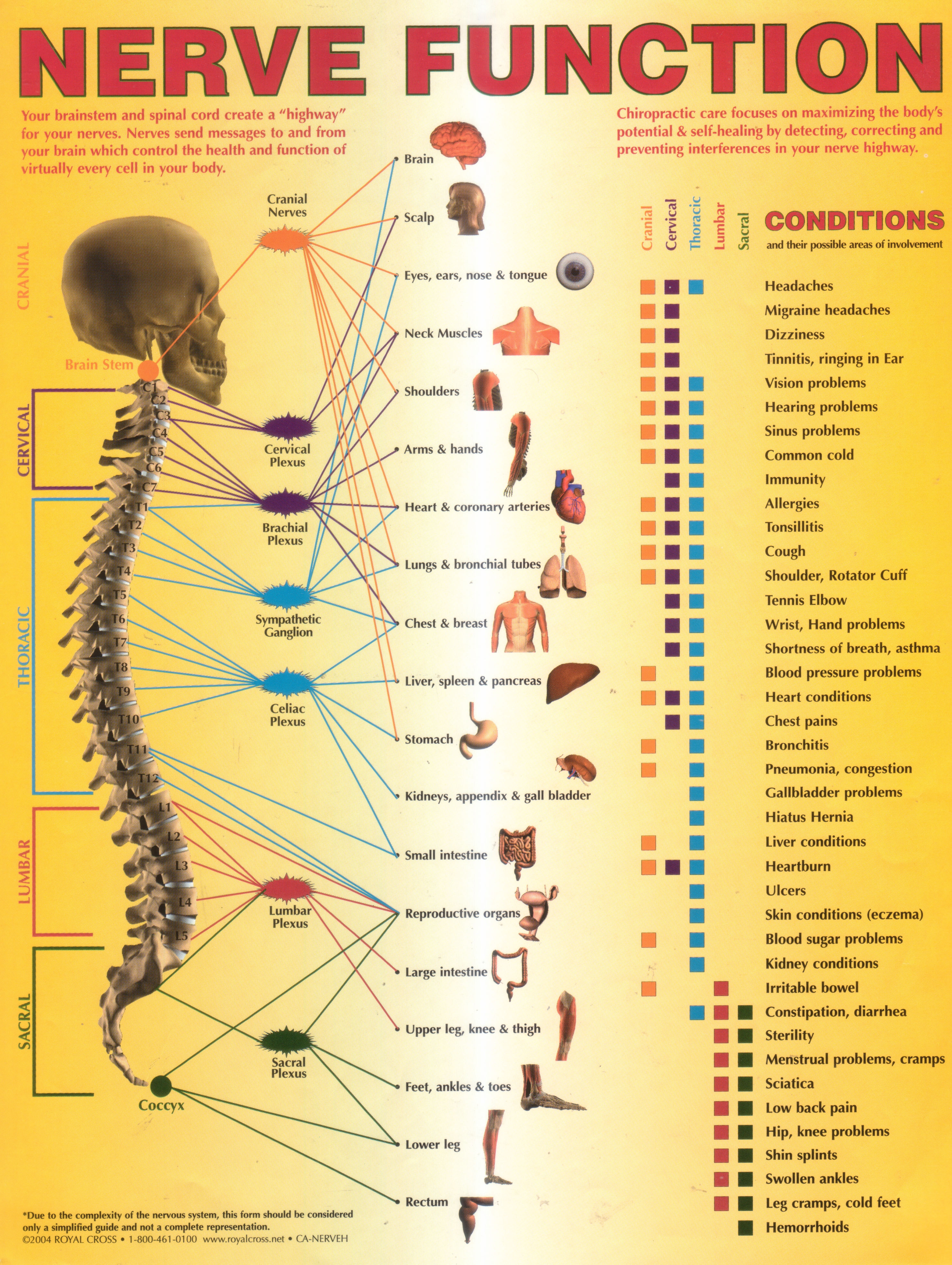
Annual World Spine Day Campaign Nerve Function Chart

Human Anatomy and Physiology Spinal Nerve Function
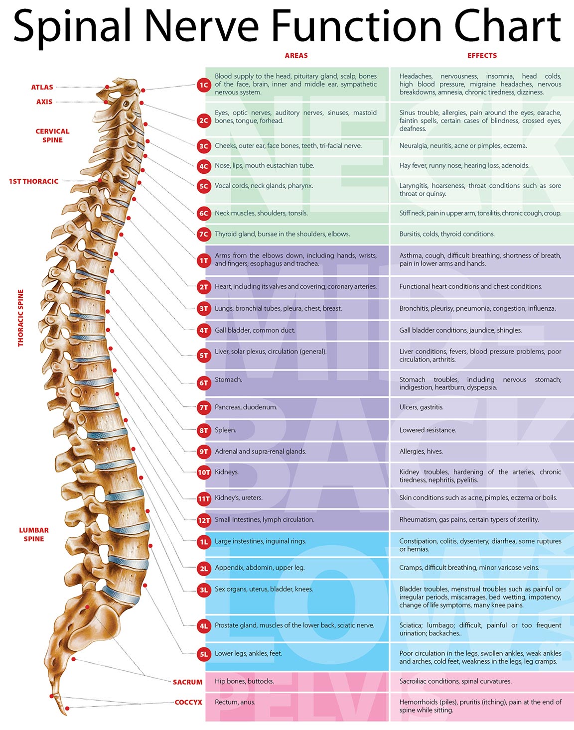
The Spinal Nerves Chart
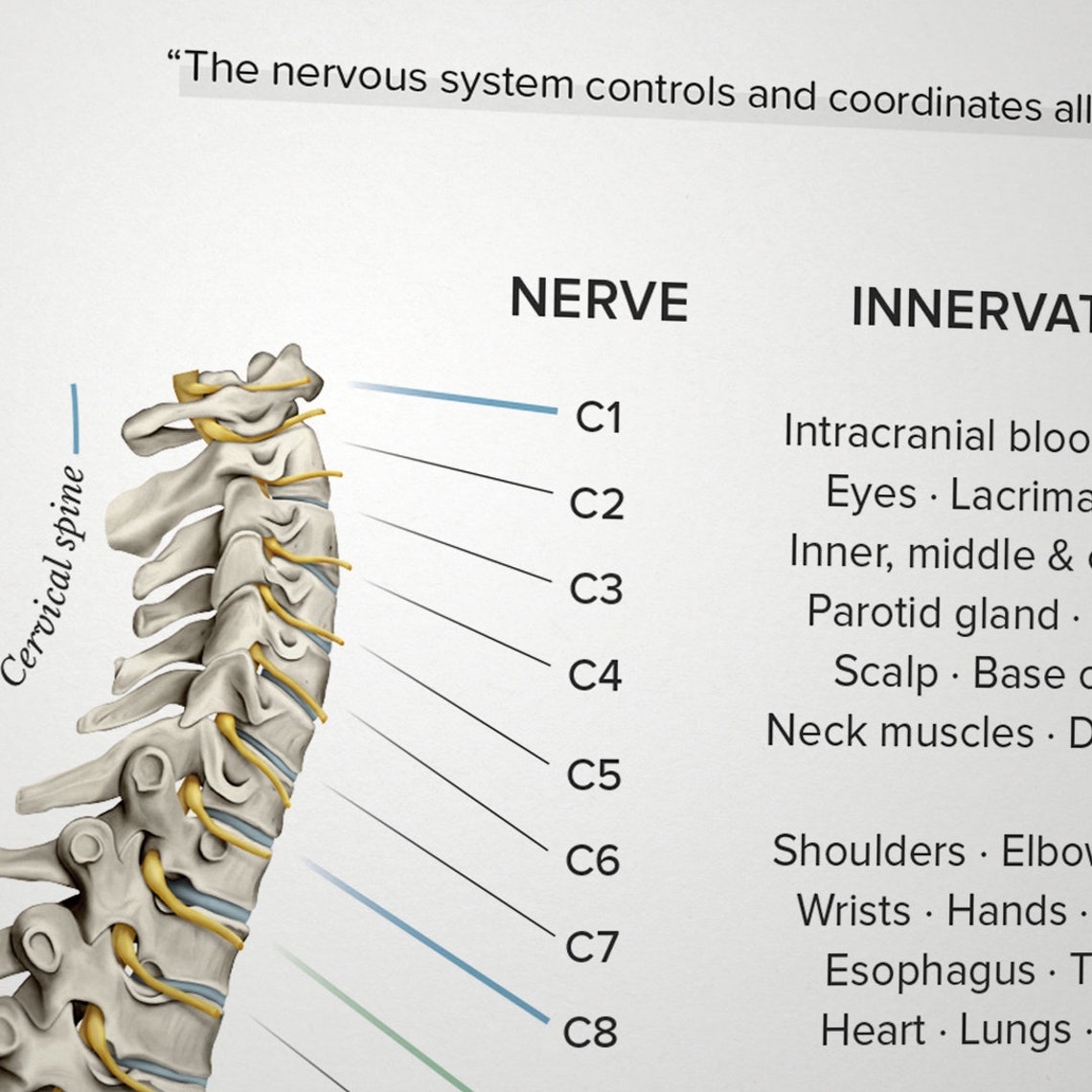
Spinal Nerve Function Chart Etsy
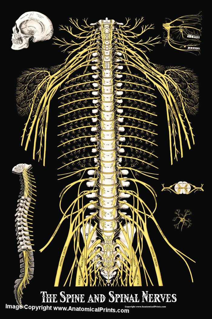
The Spine and Spinal Nerves Poster Clinical Charts and Supplies

Spinal Nerve Chart medschool doctor medicalstudent Image Credits
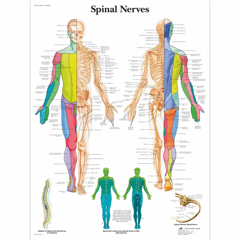
Spinal Nerves Chart MedicalSupplies.co.uk
Near The Spinal Cord Each Spinal Nerve Branches Into Two Roots.
Web Your Spinal Column Or ‘Backbone’ Is Made Up Of 24 Vertebrae:
The Point At Which A Nerve Exits The Spinal Cord Is Called A Nerve Root.
The Spinal Nerves Innervate The Skin And The Muscles Of A Specific Region.
Related Post: