Veterinary Dog Ear Cytology Chart
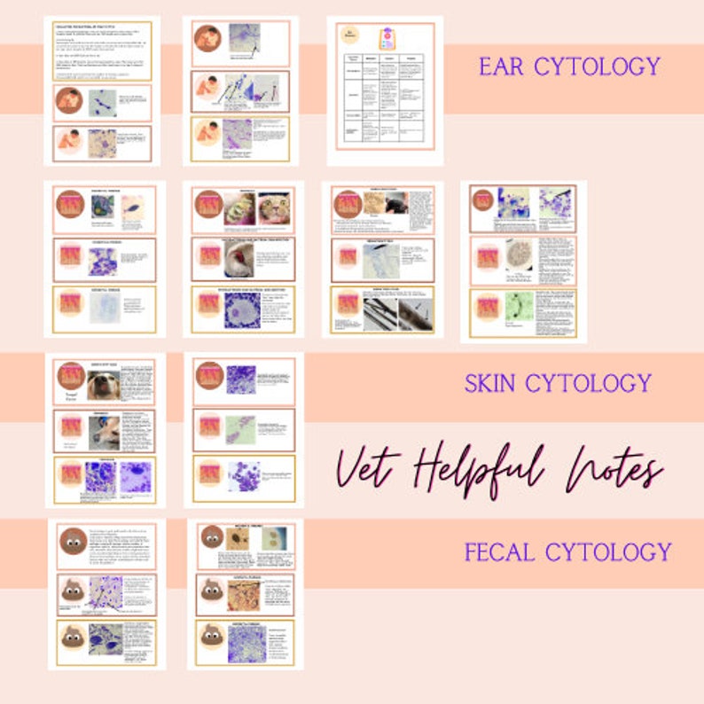
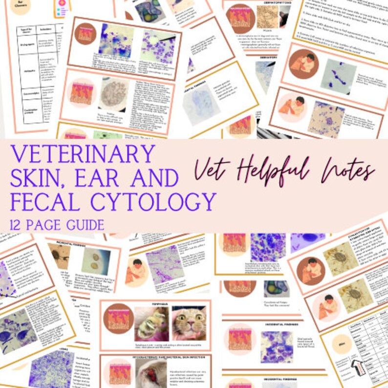
Veterinary Dog Ear Cytology Chart - Both ears should be sampled even if only 1 ear is affected in order to. Sampling, processing, and microscopic evaluation. Then, i started using pictures and videos of me actually collecting cytology in presentations and social media posts and it finally made sense. 1 restraining the patient step 1: Cytologic examination of discharge from ears should be a routine part of every ear examination. Ear cytology is a noninvasive, effective tool used to diagnose and monitor conditions of the external ear canal and pinnae. Collecting, processing, and evaluating ear. Web ear cytology method 1. This not only helps the smooth. Web ear cytology is done to identify infectious agents and can direct the vet to what treatment is best. Web this article reviews how to perform cytological examination of ear discharge, and how it can be used to characterise infection and target therapy in order to optimise. Collecting, processing, and evaluating ear. Find out how to identify bacterial, yeast and parasitic infections and how to. The slide can be air dried (pruritic material) or gently heat fixed with a. Repeat with a separate cotton bud with the opposite ear. Web veterinarians more than anything. Ear cytology is a noninvasive, effective tool used to diagnose and monitor conditions of the external ear canal and pinnae. Web ear cytology can be simple, rapid and inexpensive, and samples are relatively easy to obtain from conscious dogs. Web otitic material from each ear. The most common cells seen on ear cytology are squames,. Learn how to perform ear cytology to diagnose and monitor otitis externa in dogs and cats. Web ear cytology is done to identify infectious agents and can direct the vet to what treatment is best. Web otitic material from each ear can be spread along opposite sides of the slide.. It is important to treat ear. Learn how to perform ear cytology to diagnose and monitor otitis externa in dogs and cats. Web otitic material from each ear can be spread along opposite sides of the slide. Also, in cases of mixed infection,. This not only helps the smooth. The outer ear flap is. The most common cells seen on ear cytology are squames,. This article covers the rationale, supplies, steps, and microscopic evaluation of ear samples. Collecting, processing, and evaluating ear. With a cotton bud, gently remove some of the material from just inside the ear canal. 1 restraining the patient step 1: Both ears should be sampled even if only 1 ear is affected in order to. It is important to treat ear. This article covers the rationale, supplies, steps, and microscopic evaluation of ear samples. This not only helps the smooth. Cytology can characterize the severity of overgrowth or infection; 1 restraining the patient step 1: Repeat with a separate cotton bud with the opposite ear. Web the value of cytology exceeds simple identification of organisms. Web the veterinary nurse plays a vital role in helping the veterinary surgeon to examine the ear, take samples and perform microscopy. Collecting, processing, and evaluating ear. Web this article reviews how to perform cytological examination of ear discharge, and how it can be used to characterise infection and target therapy in order to optimise. Web veterinarians more than anything. This not only helps the smooth. Cytologic examination of discharge from ears should be a routine part of every ear examination. Web the proper diagnostic workup for canine ear disease. Collecting, processing, and evaluating ear. Web veterinarians more than anything. Web the pictures above show a diagram of the right ear as it appears if you are looking at the dog’s head from the front and a ct scan of the head. • ask an assistant to. Repeat with a separate cotton bud with the opposite ear. Web otitic material from each ear can be spread along opposite sides of the slide. Web dogs with otitis should be reevaluated with otic examination and cytology every 2 to 4 weeks, depending on severity, to assess response to therapy. Web the veterinary nurse plays a vital role in helping. Repeat with a separate cotton bud with the opposite ear. Learn how to perform ear cytology to diagnose and monitor otitis externa in dogs and cats. The slide can be air dried (pruritic material) or gently heat fixed with a hair dryer (waxy/oily. Web otitic material from each ear can be spread along opposite sides of the slide. The most common cells seen on ear cytology are squames,. Also, in cases of mixed infection,. It is important to treat ear. This not only helps the smooth. The outer ear flap is. Collecting, processing, and evaluating ear. Web veterinarians more than anything. • ask an assistant to. Web learn how to collect and interpret ear cytology samples in dogs with otitis externa. Web the proper diagnostic workup for canine ear disease. Cytology can characterize the severity of overgrowth or infection; Ear cytology is a noninvasive, effective tool used to diagnose and monitor conditions of the external ear canal and pinnae.
Animal Ear Cytology Identification Chart

Canine Ear Cytology Chart

Idexx Ear Cytology Chart

Animal Ear Cytology Identification Chart

Vet Tech Skin and Ear Cytology Notes Vet Tech and Vet Nurse Etsy

Dog Ear Cytology Rods

Canine Ear Cytology Chart
Veterinary Dog Ear Cytology Chart

Vet Tech Skin and Ear Cytology Notes Vet Tech and Vet Nurse Etsy

Ear Cytology Dog Chart
Find Out How To Identify Bacterial, Yeast And Parasitic Infections And How To.
Web The Pictures Above Show A Diagram Of The Right Ear As It Appears If You Are Looking At The Dog’s Head From The Front And A Ct Scan Of The Head.
With A Cotton Bud, Gently Remove Some Of The Material From Just Inside The Ear Canal.
Web Ear Cytology Is Done To Identify Infectious Agents And Can Direct The Vet To What Treatment Is Best.
Related Post:
