Visual Field Defects Chart
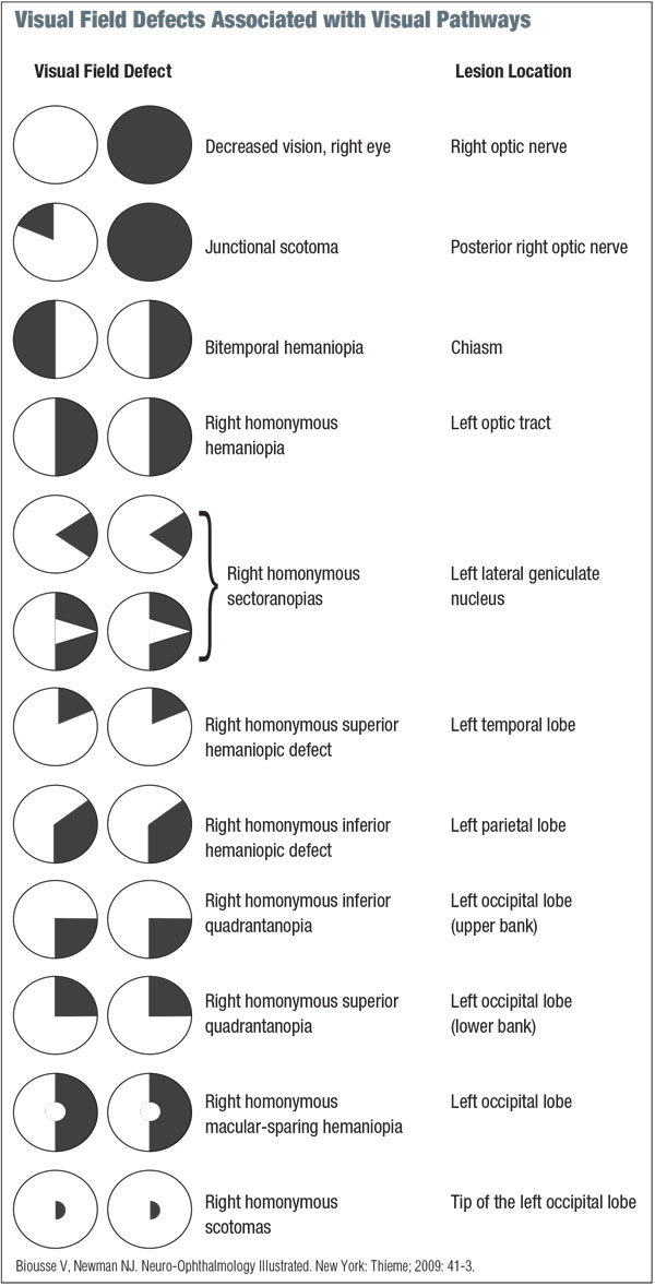
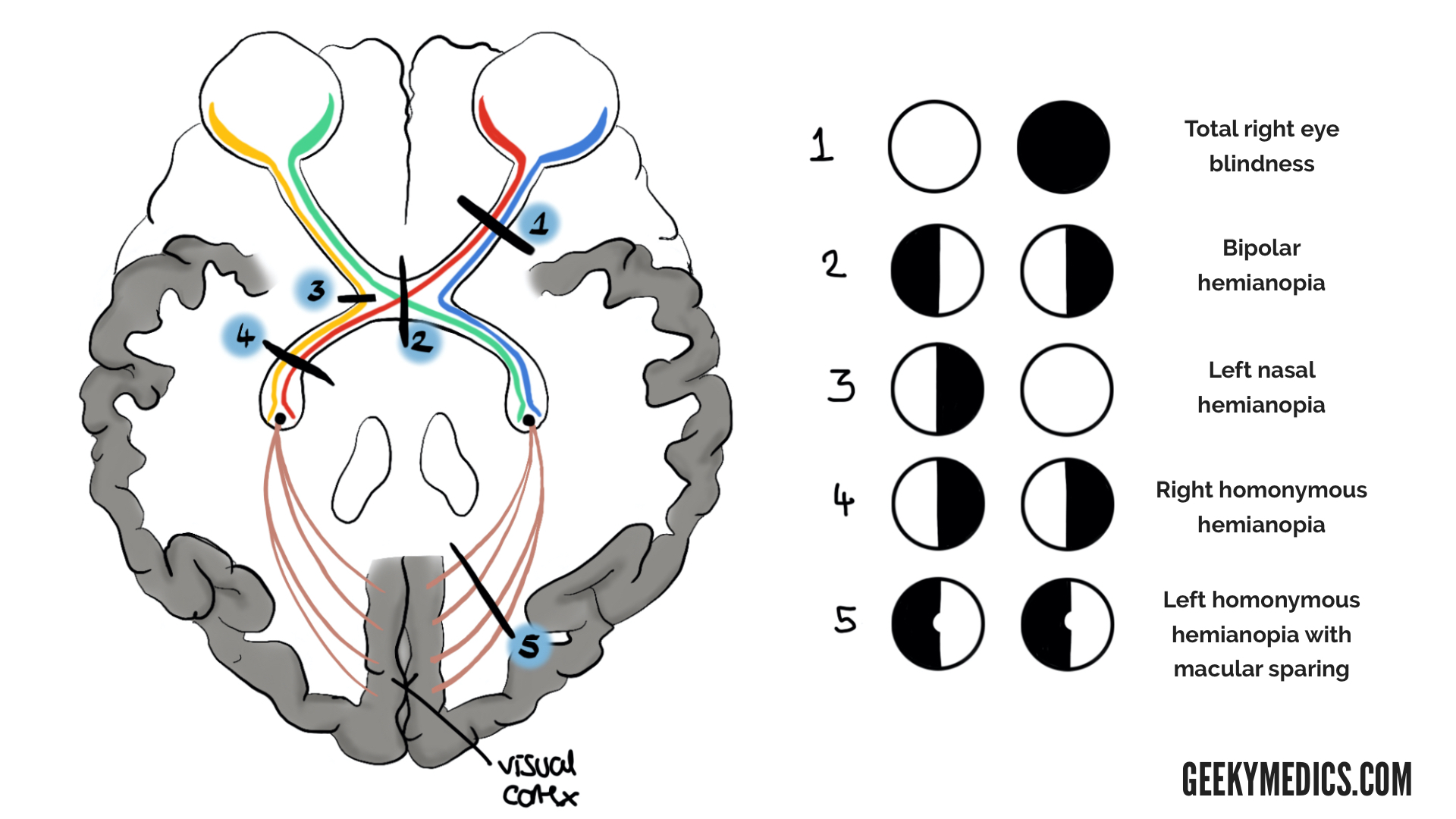
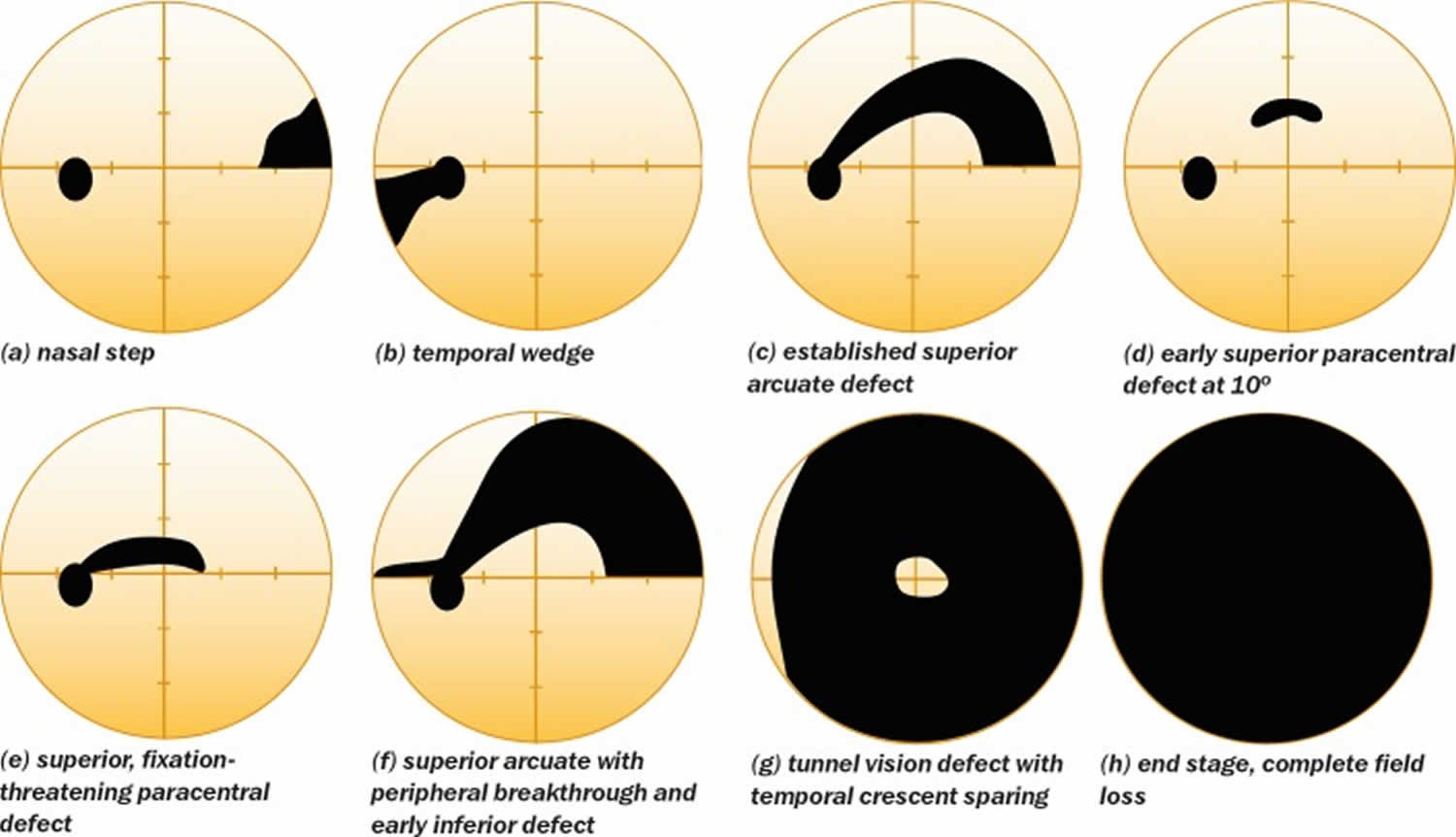
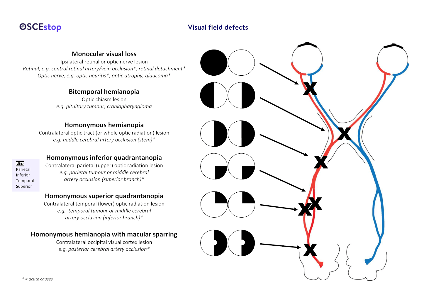
Visual Field Defects Chart - Lesions compressing the chiasm, such as pituitary adenomas, therefore cause bitemporal hemianopia. They are caused by lesions along the visual pathway, which stretches from the retina to the visual cortex in the brain. Web this chapter explores how the organization of the eye and visual system dictates specific, recognizable patterns of visual field loss in disease. Our normal field of vision is typically 135º vertically and 180º horizontally (160º for monocular vision). Compare to the previous visual fields. Does not cross the horizontal median. Web look at the pattern. Web is there a significant defect? Web damage to visual mechanisms along various portions of the visual pathways from the optics and photoreceptors up to the visual centers of the brain will produce different shapes and patterns of visual field loss. False negatives are identified when the patient does not respond to a light stimulus that should have been detected, based upon earlier responses. Does not cross the horizontal median Web is there a significant defect? Web visual field defects. The law requires that all. Look for a cluster of two or more defects, and look at the summary data, including mean deviation, pattern standard deviation, visual field index (vfi) and the glaucoma hemifield test. They’re drawn as if you are the pt! False negatives are identified when the patient does not respond to a light stimulus that should have been detected, based upon earlier responses. Learn about the top 5 most common fields! Our normal field of vision is typically 135º vertically and 180º horizontally (160º for monocular vision). Web the blindspot is represented. They are caused by lesions along the visual pathway, which stretches from the retina to the visual cortex in the brain. Web field defects are more common in individuals with superficially located or visible disc drusens. Web visual field defects. Web early (or even moderate) visual field defects often go unnoticed, particularly if only one eye is affected. The visual. Web field defects are more common in individuals with superficially located or visible disc drusens. Results must be interpreted critically (reliability and repeatability) and in conjunction with other clinical signs, symptoms and examination findings. Normal blind spot is on the retina where the optic nerve is due to no photoreceptors and is not noticeable. Causes loss of central vision. Web. 1 2 skilled interpretation of visual field tests requires a good grasp and application of this prior knowledge. The visual field describes the area that can be seen by an individual with their eyes fixed on a single point. Web field defects are more common in individuals with superficially located or visible disc drusens. Measured in degrees from fixation, how. Ischemic optic neuropathy, hemibranch retinal artery occlusion, retinal detachment. Results must be interpreted critically (reliability and repeatability) and in conjunction with other clinical signs, symptoms and examination findings. Learn about the top 5 most common fields! Lesions compressing the chiasm, such as pituitary adenomas, therefore cause bitemporal hemianopia. The visual field test is among the most important tests to learn. Our normal field of vision is typically 135º vertically and 180º horizontally (160º for monocular vision). Some more common ones are included here. Central field loss results from degeneration of the fovea and occurs with: Look for a cluster of two or more defects, and look at the summary data, including mean deviation, pattern standard deviation, visual field index (vfi). Get ready for easy referencing! Compare to the previous visual fields. They are caused by lesions along the visual pathway, which stretches from the retina to the visual cortex in the brain. The left eye has inferior field loss, and the right eye has superior field loss. Web some common types of visual field defects and their more common differentials. Central scotoma is due to lesion of macula. Web visual field defects are, therefore, not limited to glaucoma. Visual field defects are a partial loss of the regular field of vision. Web this chapter explores how the organization of the eye and visual system dictates specific, recognizable patterns of visual field loss in disease. False negatives are identified when the. Web is there a significant defect? Instead we recommend two excellent recent reviews. There are many causes of visual field loss. Web early (or even moderate) visual field defects often go unnoticed, particularly if only one eye is affected. Get ready for easy referencing! The left eye has inferior field loss, and the right eye has superior field loss. Normal blind spot is on the retina where the optic nerve is due to no photoreceptors and is not noticeable. Useful aspects of eye anatomy. Web visual field defects. What are visual field defects? The visual field test is among the most important tests to learn to interpret as you begin your career in ophthalmology. Web damage to visual mechanisms along various portions of the visual pathways from the optics and photoreceptors up to the visual centers of the brain will produce different shapes and patterns of visual field loss. Our normal field of vision is typically 135º vertically and 180º horizontally (160º for monocular vision). Compare to the previous visual fields. The images in figure 2 represent what a scene may look like to someone with different visual field defects in each eye. A visual field defect is a loss of part of the usual field of vision. Causes loss of central vision. Learn about the top 5 most common fields! To assist you in being able to properly interpret visual fields, a table indicating the classic patterns of visual field loss. There are many causes of visual field loss. Web it is beyond the scope of this paper to cover the neuroanatomical localisation of visual field defects.
How to Describe Visual Field Defects
Visual field defect American Academy of Ophthalmology

Image result for visual field defects and light reflex chart Chart

Different Types Of Visual Field Defects BEST GAMES WALKTHROUGH

Visual Field Defects Ophthalmology Medbullets Step 2/3

Dx Schema Visual Field Defects The Clinical Problem Solvers

Visual Field Defects on Meducation Optometry education, Optometry

Visual Field Defects Geeky Medics

Visual field test, visual field test results interpretation

Visual field defects OSCEstop OSCE Learning
At The Optic Chiasm, Fibres From The Nasal Half Of The Retina, Corresponding To The Temporal Visual Field, Decussate.
Web The Field May Often Look Normal Or “Cleaner” (With Fewer Defects Depicted) On Patients With Values Of 5% To 10% Or Higher.
Lesions Compressing The Chiasm, Such As Pituitary Adenomas, Therefore Cause Bitemporal Hemianopia.
It Is Very Important To Examine The Retina And Optic Disc Carefully To Assess Whether Or Not A Visual Field Defect Matches The Appearance Of The Disc And Retina, Or Fits With Other Clinical Signs.
Related Post: