Mucous Membrane Color Chart Dogs
Mucous Membrane Color Chart Dogs - 1 normal, healthy canine mucous membranes are pink; Anaphylaxis is defined as the acute onset of a hypersensitivity reaction causing the release of mediators from mast cells and basophils. The tissue in the mucous membranes is very thin and well supplied with blood vessels, so changes within body tissue are more visible in the mucous membranes than elsewhere in the body. Web mucous membranes is a fancy way to say gum color and should be the color of “bubble gum” pink on a regular basis. The most common colorimetric scale in use in veterinary medicine is the faffa malan chart (famacha), which is designed to Pallor or hyperemia are suggestive of disruptions to tissue perfusion or oxygenation status. Triage is the art of assigning priority to emergency patients and their problems based on rapid assessment of historical and physical parameters ( see table: For instance, the cyanotic patient is experiencing severe hypoxemia. Derby of river road animal hospital and learn how to check your dog's gum color, otherwise known as mucous membrane. You may notice other colors, such as blue or white gums, which indicate an emergency. Anaphylaxis is defined as the acute onset of a hypersensitivity reaction causing the release of mediators from mast cells and basophils. The present study revealed the efficacy of nsaids, fluid. Web white or pale mucous membranes typically indicate vasoconstriction or anemia. For instance, the cyanotic patient is experiencing severe hypoxemia. Web normal mucous membranes, dog. The color of the mucous membranes can tell you a lot about your dog's health. Web mucous membrane colour reflects the oxygenation and perfusion (flow of blood through the body’s blood vessels) of the tissues1. A resolution of an increased blood lactate to 2. Web locations to assess mucous membranes in dogs include the gingival, conjunctival, vulvar, and penile mucosa.. Web images / normal mucous membranes, dog. A resolution of an increased blood lactate to 2. Learn what is your dog's normal gum color. Web abnormal mucous membrane colors include pale pink, white, yellow, blue, chocolate brown, red, and “cherry red.” these color changes represent critical physical examination findings that facilitate the diagnostic plan for every patient. When the mucous. When the mucous membranes become pale or discolored, this can be a sign of a serious medical condition. Pallor or hyperemia are suggestive of disruptions to tissue perfusion or oxygenation status. Web abnormal mucous membrane colors include pale pink, white, yellow, blue, chocolate brown, red, and “cherry red.” these color changes represent critical physical examination findings that facilitate the diagnostic. You may notice other colors, such as blue or white gums, which indicate an emergency. The tissue in the mucous membranes is very thin and well supplied with blood vessels, so changes within body tissue are more visible in the mucous membranes than elsewhere in the body. Web in most healthy dogs, the mucous membranes are pink and moist. If. Your pet's mucous membrane colour can be an indication of cardiovascular and respiratory health and your vet may ask you to check this. Both of these conditions may lead to decreased oxygen delivery to the tissues. Anaphylaxis is defined as the acute onset of a hypersensitivity reaction causing the release of mediators from mast cells and basophils. Mucous membranes are. The following is a list of the different colors you may witness and what each of those colors might indicate. 307 views 3 years ago river road animal hospital. Web at animalwised, we look at pale mucus membranes in dogs. Triage is the art of assigning priority to emergency patients and their problems based on rapid assessment of historical and. Web normal mucous membranes, dog. Colorimetric scales have been used in both human and veterinary medicine to assess mucosal color. Triage is the art of assigning priority to emergency patients and their problems based on rapid assessment of historical and physical parameters ( see table: Web at animalwised, we look at pale mucus membranes in dogs. Increased serum bilirubin due. Often, a specific cause for anaphylaxis is not known. Web white or pale mucous membranes typically indicate vasoconstriction or anemia. Vasoconstriction is usually a result of hypovolemic or cardiogenic shock. Web normal vital signs include taking measurements and observations of the heart rate, respiratory rate, temperature, capillary refill time and mucous membrane colour and hydration status. For instance, the cyanotic. Increased serum bilirubin due to hepatic disease or hemolysis. Web mucous membrane color is determined by the presence of oxygenated hemoglobin in capillaries and vessels just beneath the mucosal surface. We understand what dog mucus membrane discoloration can mean, as well as provide some other symptoms to take into account. Pallor or hyperemia are suggestive of disruptions to tissue perfusion. Pallor or hyperemia are suggestive of disruptions to tissue perfusion or oxygenation status. 307 views 3 years ago river road animal hospital. Often, a specific cause for anaphylaxis is not known. Both of these conditions may lead to decreased oxygen delivery to the tissues. Web in a healthy pet, the mucous membranes are pink and moist. The present study revealed the efficacy of nsaids, fluid. Web mucous membrane color is determined by the presence of oxygenated hemoglobin in capillaries and vessels just beneath the mucosal surface. Severe hypoxemia or decompensatory shock. Normal pcv and adequate perfusion. Web the endpoints typically reflect perfusion status and include heart rate, blood pressure, central venous pressure, mucous membrane color, capillary refill time, and pulse intensity. Vasoconstriction is usually a result of hypovolemic or cardiogenic shock. You may notice other colors, such as blue or white gums, which indicate an emergency. We understand what dog mucus membrane discoloration can mean, as well as provide some other symptoms to take into account. Web cyanotic mucous membranes, dog. The tissue in the mucous membranes is very thin and well supplied with blood vessels, so changes within body tissue are more visible in the mucous membranes than elsewhere in the body. Web abnormal mucous membrane colors include pale pink, white, yellow, blue, chocolate brown, red, and “cherry red.” these color changes represent critical physical examination findings that facilitate the diagnostic plan for every patient.
Bad breath Thick, tacky saliva Dry cracked tongue Plaque & calculus

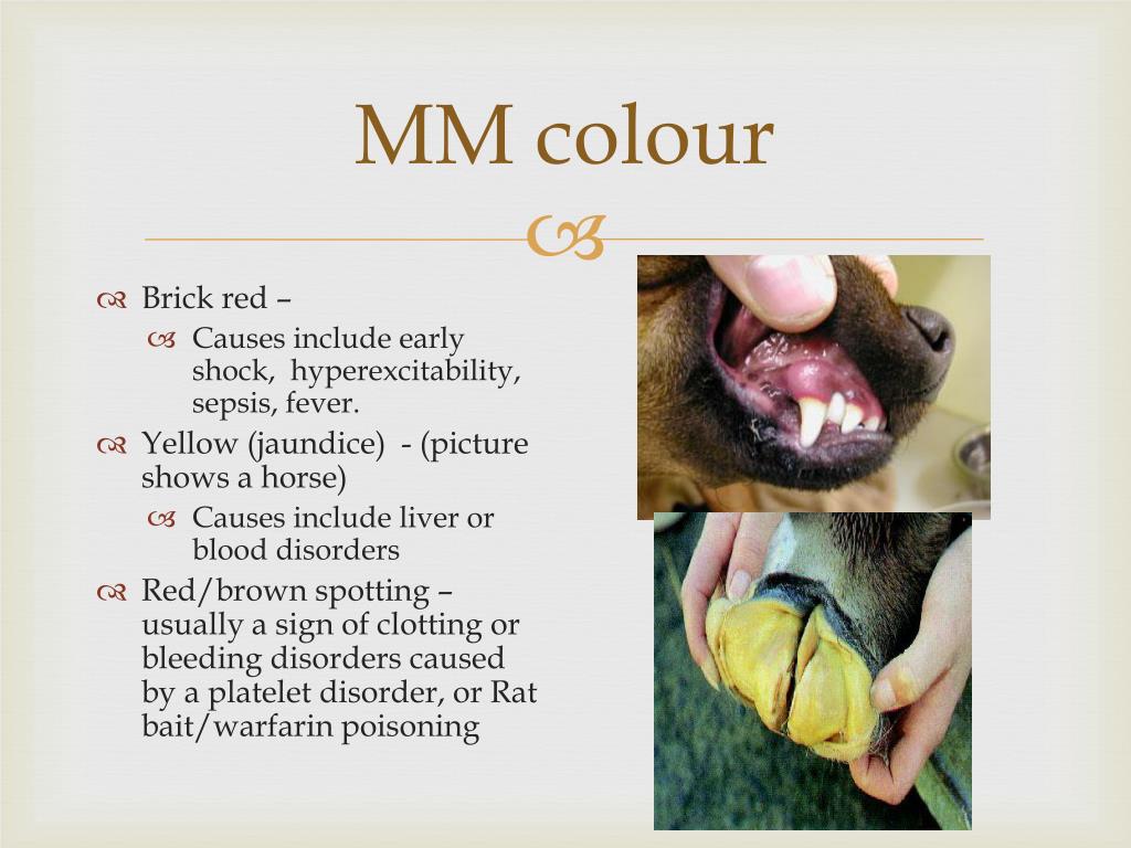
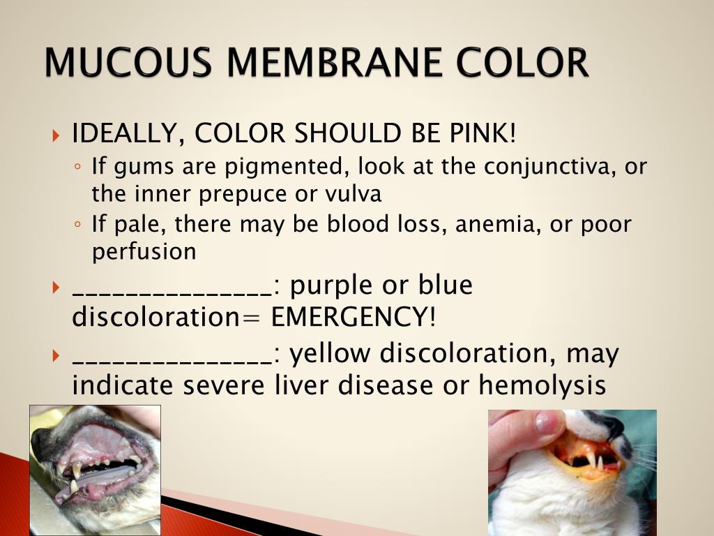
PPT Clinical Examination of Animals PowerPoint Presentation, free

Mucous Membrane Color Chart Dogs A Visual Reference of Charts Chart

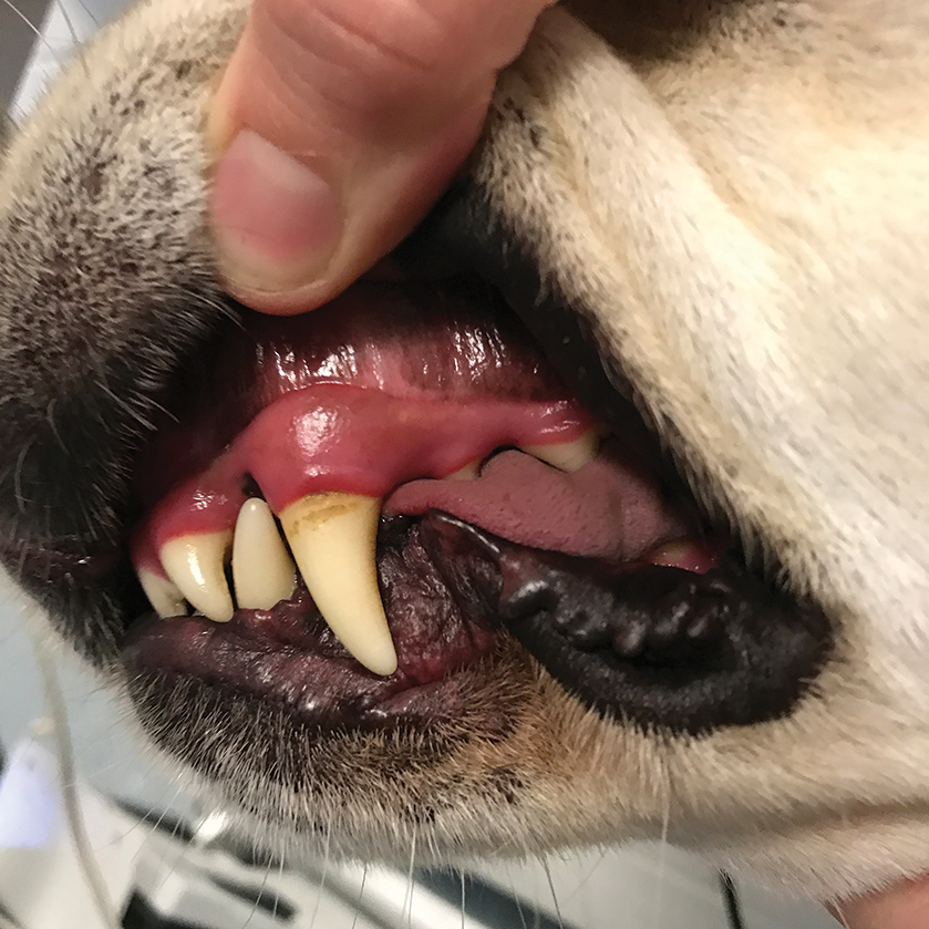
What Do Healthy Dog Gums Look Like? PuppyLists

ammaarahxyburrows51a how to check your dog or cats blood circulation

Checking your pet's mucous membrane colour YouTube

Changes in Mucous Membrane Color Dog Symptoms

PPT Clinical Examination of Animals PowerPoint Presentation, free

Mucous Membrane Color in Dogs Pet Friendly House

PPT Clinical Examination of Animals PowerPoint Presentation, free
Web Mucous Membrane Colour Reflects The Oxygenation And Perfusion (Flow Of Blood Through The Body’s Blood Vessels) Of The Tissues1.
Both Of These Conditions May Lead To Decreased Oxygen Delivery To The Tissues.
Web In The Author’s Experience, Mucous Membrane Color May Vary Among Patients, As Some Canine Breeds Such As Weimaraners And Red Doberman Pinschers Have Naturally Pinker Membranes.
For Instance, The Cyanotic Patient Is Experiencing Severe Hypoxemia.
Related Post: